de Lima S, Mietto B S, Paula C, Muniz T, Martinez A M B, Gardino P F
Laboratório de Neurobiologia da Retina, Centro de Ciências da Saúde, Instituto de Biofísica Carlos Chagas Filho, Rio de Janeiro, RJ, Brasil.
Laboratório de Neurodegeneração e Reparo, Departamento de Patologia, Faculdade de Medicina, Centro de Ciências da Saúde, Universidade Federal do Rio de Janeiro, Rio de Janeiro, RJ, Brasil.
Braz J Med Biol Res. 2016;49(4):e5106. doi: 10.1590/1414-431X20155106. Epub 2016 Mar 18.
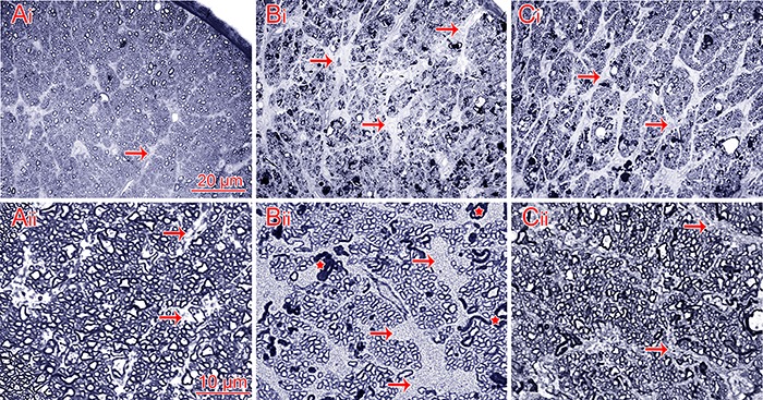
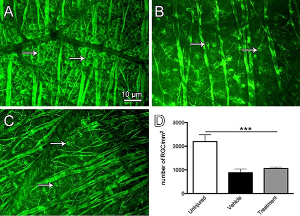
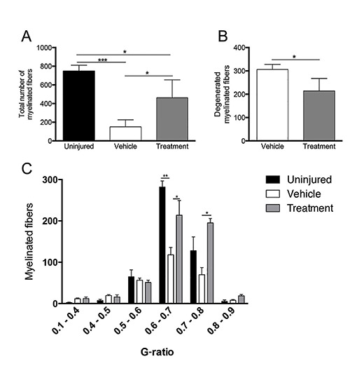
After a traumatic injury to the central nervous system, the distal stumps of axons undergo Wallerian degeneration (WD), an event that comprises cytoskeleton and myelin breakdown, astrocytic gliosis, and overexpression of proteins that inhibit axonal regrowth. By contrast, injured neuronal cell bodies show features characteristic of attempts to initiate the regenerative process of elongating their axons. The main molecular event that leads to WD is an increase in the intracellular calcium concentration, which activates calpains, calcium-dependent proteases that degrade cytoskeleton proteins. The aim of our study was to investigate whether preventing axonal degeneration would impact the survival of retinal ganglion cells (RGCs) after crushing the optic nerve. We observed that male Wistar rats (weighing 200-400 g; n=18) treated with an exogenous calpain inhibitor (20 mM) administered via direct application of the inhibitor embedded within the copolymer resin Evlax immediately following optic nerve crush showed a delay in the onset of WD. This delayed onset was characterized by a decrease in the number of degenerated fibers (P<0.05) and an increase in the number of preserved fibers (P<0.05) 4 days after injury. Additionally, most preserved fibers showed a normal G-ratio. These results indicated that calpain inhibition prevented the degeneration of optic nerve fibers, rescuing axons from the process of axonal degeneration. However, analysis of retinal ganglion cell survival demonstrated no difference between the calpain inhibitor- and vehicle-treated groups, suggesting that although the calpain inhibitor prevented axonal degeneration, it had no effect on RGC survival after optic nerve damage.
中枢神经系统遭受创伤性损伤后,轴突的远端残端会发生沃勒变性(WD),这一过程包括细胞骨架和髓鞘分解、星形胶质细胞增生以及抑制轴突再生的蛋白质过度表达。相比之下,受损的神经元细胞体表现出试图启动轴突伸长再生过程的特征。导致沃勒变性的主要分子事件是细胞内钙浓度升高,这会激活钙蛋白酶,即降解细胞骨架蛋白的钙依赖性蛋白酶。我们研究的目的是探讨预防轴突变性是否会对视神经挤压后视网膜神经节细胞(RGCs)的存活产生影响。我们观察到,在视神经挤压后立即通过直接应用包埋在共聚物树脂Evlax中的外源性钙蛋白酶抑制剂(20 mM)处理的雄性Wistar大鼠(体重200 - 400 g;n = 18),沃勒变性的起始出现延迟。这种延迟起始的特征是损伤后4天变性纤维数量减少(P < 0.05),保留纤维数量增加(P < 0.05)。此外,大多数保留纤维显示出正常的G比值。这些结果表明,抑制钙蛋白酶可防止视神经纤维变性,使轴突免于轴突变性过程。然而,视网膜神经节细胞存活分析表明,钙蛋白酶抑制剂处理组和溶剂处理组之间没有差异,这表明尽管钙蛋白酶抑制剂可防止轴突变性,但对视神经损伤后的视网膜神经节细胞存活没有影响。