• 文献检索
  • 文档翻译
  • 深度研究
  • 学术资讯
  • Suppr Zotero 插件Zotero 插件
  • 邀请有礼
  • 套餐&价格
  • 历史记录
应用&插件
Suppr Zotero 插件Zotero 插件浏览器插件Mac 客户端Windows 客户端微信小程序
定价
高级版会员购买积分包购买API积分包
服务
文献检索文档翻译深度研究API 文档MCP 服务
关于我们
关于 Suppr公司介绍联系我们用户协议隐私条款
关注我们

Suppr 超能文献

核心技术专利:CN118964589B侵权必究
粤ICP备2023148730 号-1Suppr @ 2026

文献检索

告别复杂PubMed语法,用中文像聊天一样搜索,搜遍4000万医学文献。AI智能推荐,让科研检索更轻松。

立即免费搜索

文件翻译

保留排版,准确专业,支持PDF/Word/PPT等文件格式,支持 12+语言互译。

免费翻译文档

深度研究

AI帮你快速写综述,25分钟生成高质量综述,智能提取关键信息,辅助科研写作。

立即免费体验

使用原子力显微镜区分正常细胞和癌细胞

Discrimination Between Normal and Cancerous Cells Using AFM.

作者信息

Lekka Małgorzata

机构信息

Institute of Nuclear Physics, PAS, Radzikowskiego 152, 31-342 Kraków, Poland.

出版信息

Bionanoscience. 2016;6:65-80. doi: 10.1007/s12668-016-0191-3. Epub 2016 Jan 30.

DOI:10.1007/s12668-016-0191-3
PMID:27014560
原文链接:https://pmc.ncbi.nlm.nih.gov/articles/PMC4778153/
Abstract

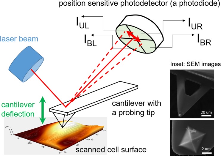
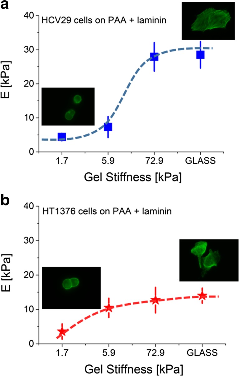
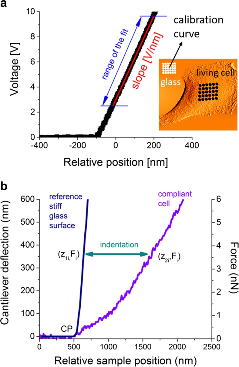
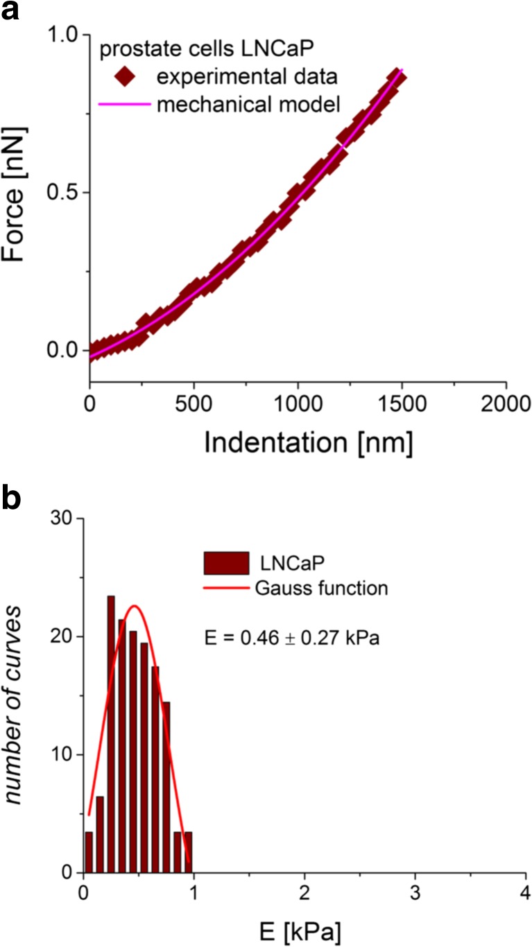
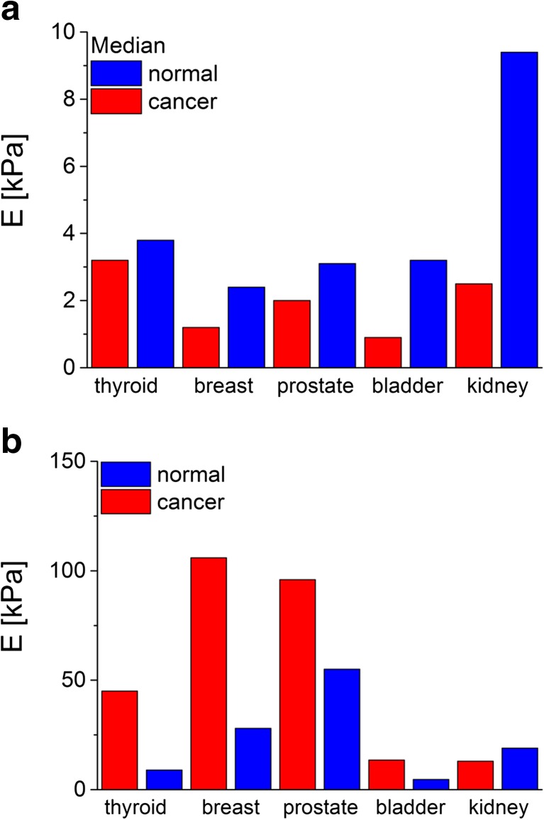
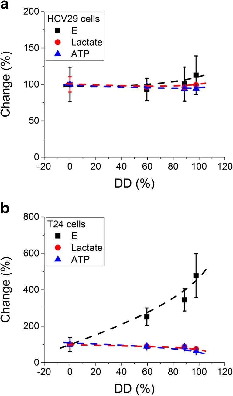
Currently, biomechanics of living cells is in the focus of interest due to noticeable capability of such techniques like atomic force microscopy (AFM) to probe cellular properties at the single cell level directly on living cells. The research carried out, so far, delivered data showing, on the one hand, the use of cellular mechanics as a biomarker of various pathological changes, which, on the other hand, reveal relative nature of biomechanics. In the AFM, the elastic properties of living cells are delivered from indentation experiments and described quantitatively by Young's modulus defined here as a measure of cellular deformability. Here, the AFM studies directly comparing the mechanical properties of normal and cancerous cells are summarized and presented together with a few important issues related to the relativeness of Young's modulus.

摘要

目前,活细胞生物力学成为研究热点,这是因为诸如原子力显微镜(AFM)等技术具有显著能力,能够在活细胞上直接于单细胞水平探测细胞特性。迄今为止所开展的研究提供的数据表明,一方面,细胞力学可作为各种病理变化的生物标志物,另一方面,这些数据也揭示了生物力学的相对性质。在原子力显微镜中,活细胞的弹性特性通过压痕实验得出,并由杨氏模量进行定量描述,在此将杨氏模量定义为细胞可变形性的一种度量。本文总结了直接比较正常细胞和癌细胞力学特性的原子力显微镜研究,并阐述了与杨氏模量相对性相关的一些重要问题。

https://cdn.ncbi.nlm.nih.gov/pmc/blobs/1ed6/4778153/6b52df67856b/12668_2016_191_Fig10_HTML.jpg
https://cdn.ncbi.nlm.nih.gov/pmc/blobs/1ed6/4778153/839527797ec4/12668_2016_191_Fig1_HTML.jpg
https://cdn.ncbi.nlm.nih.gov/pmc/blobs/1ed6/4778153/420e964134fb/12668_2016_191_Fig2_HTML.jpg
https://cdn.ncbi.nlm.nih.gov/pmc/blobs/1ed6/4778153/c731156d5379/12668_2016_191_Fig3_HTML.jpg
https://cdn.ncbi.nlm.nih.gov/pmc/blobs/1ed6/4778153/be65caa3717e/12668_2016_191_Fig4_HTML.jpg
https://cdn.ncbi.nlm.nih.gov/pmc/blobs/1ed6/4778153/8e754b28f43c/12668_2016_191_Fig5_HTML.jpg
https://cdn.ncbi.nlm.nih.gov/pmc/blobs/1ed6/4778153/a56e8d20045d/12668_2016_191_Fig6_HTML.jpg
https://cdn.ncbi.nlm.nih.gov/pmc/blobs/1ed6/4778153/bb871e6c754e/12668_2016_191_Fig7_HTML.jpg
https://cdn.ncbi.nlm.nih.gov/pmc/blobs/1ed6/4778153/e4b33273a7e3/12668_2016_191_Fig8_HTML.jpg
https://cdn.ncbi.nlm.nih.gov/pmc/blobs/1ed6/4778153/aa4b6cb41f5b/12668_2016_191_Fig9_HTML.jpg
https://cdn.ncbi.nlm.nih.gov/pmc/blobs/1ed6/4778153/6b52df67856b/12668_2016_191_Fig10_HTML.jpg

相似文献

1
Discrimination Between Normal and Cancerous Cells Using AFM.使用原子力显微镜区分正常细胞和癌细胞
Bionanoscience. 2016;6:65-80. doi: 10.1007/s12668-016-0191-3. Epub 2016 Jan 30.
2
Cancer cell recognition--mechanical phenotype.癌细胞识别——力学表型。
Micron. 2012 Dec;43(12):1259-66. doi: 10.1016/j.micron.2012.01.019. Epub 2012 Feb 11.
3
Measuring Elastic Properties of Single Cancer Cells by AFM.通过原子力显微镜测量单个癌细胞的弹性特性。
Methods Mol Biol. 2019;1886:315-324. doi: 10.1007/978-1-4939-8894-5_18.
4
Atomic force microscopy studies on cellular elastic and viscoelastic properties.原子力显微镜研究细胞的弹性和粘弹性特性。
Sci China Life Sci. 2018 Jan;61(1):57-67. doi: 10.1007/s11427-016-9041-9. Epub 2017 Jun 29.
5
Investigation into local cell mechanics by atomic force microscopy mapping and optical tweezer vertical indentation.通过原子力显微镜测绘和光镊垂直压痕研究局部细胞力学。
Nanotechnology. 2016 Feb 12;27(6):065102. doi: 10.1088/0957-4484/27/6/065102. Epub 2015 Dec 18.
6
Applicability of atomic force microscopy to determine cancer-related changes in cells.原子力显微镜在确定细胞癌变相关变化中的适用性。
Philos Trans A Math Phys Eng Sci. 2022 Sep 19;380(2232):20210346. doi: 10.1098/rsta.2021.0346. Epub 2022 Aug 1.
7
Experimental extraction of Young's modulus of MCF-7 tissue using atomic force microscopy and the spherical contact models.使用原子力显微镜和球形接触模型对MCF-7组织杨氏模量进行实验提取。
Eur Biophys J. 2023 Feb;52(1-2):81-90. doi: 10.1007/s00249-023-01642-3. Epub 2023 Mar 16.
8
Robust strategies for automated AFM force curve analysis--I. Non-adhesive indentation of soft, inhomogeneous materials.用于自动原子力显微镜力曲线分析的稳健策略——I. 软质非均匀材料的非粘性压痕
J Biomech Eng. 2007 Jun;129(3):430-40. doi: 10.1115/1.2720924.
9
Micropipette-assisted atomic force microscopy for single-cell 3D manipulations and nanomechanical measurements.微移液器辅助原子力显微镜用于单细胞 3D 操作和纳米力学测量。
Nanoscale. 2023 Aug 17;15(32):13346-13358. doi: 10.1039/d3nr02404k.
10
Quantitative study of the elastic modulus of loosely attached cells in AFM indentation experiments.原子力显微镜压痕实验中松动附着细胞的弹性模量的定量研究。
Biophys J. 2013 May 21;104(10):2123-31. doi: 10.1016/j.bpj.2013.04.019.

引用本文的文献

1
Viscoelastic recovery times of chondrocytes measured using a novel 3D-printed microfluidic device.使用新型3D打印微流控装置测量软骨细胞的粘弹性恢复时间。
Meas Sci Technol. 2025 Aug 31;36(8):085701. doi: 10.1088/1361-6501/adf65a. Epub 2025 Aug 12.
2
Effects of fluid shear stress duration on the mechanical properties of HeLa cells using atomic force microscopy.利用原子力显微镜研究流体剪切应力作用时间对HeLa细胞力学性能的影响。
PLoS One. 2025 May 5;20(5):e0321296. doi: 10.1371/journal.pone.0321296. eCollection 2025.
3
Softness or Stiffness What Contributes to Cancer and Cancer Metastasis?

本文引用的文献

1
Soft biological materials and their impact on cell function.柔软生物材料及其对细胞功能的影响。
Soft Matter. 2007 Feb 14;3(3):299-306. doi: 10.1039/b610522j.
2
Investigation into local cell mechanics by atomic force microscopy mapping and optical tweezer vertical indentation.通过原子力显微镜测绘和光镊垂直压痕研究局部细胞力学。
Nanotechnology. 2016 Feb 12;27(6):065102. doi: 10.1088/0957-4484/27/6/065102. Epub 2015 Dec 18.
3
Discrimination Between Cervical Cancer Cells and Normal Cervical Cells Based on Longitudinal Elasticity Using Atomic Force Microscopy.
柔软还是坚硬?是什么导致了癌症及癌症转移?
Cells. 2025 Apr 12;14(8):584. doi: 10.3390/cells14080584.
4
Effect of elastic modulus of tumour and non-tumour cells on vibration-induced behaviours.肿瘤细胞和非肿瘤细胞的弹性模量对振动诱导行为的影响。
Sci Rep. 2025 Apr 16;15(1):13199. doi: 10.1038/s41598-025-97837-z.
5
Single-cell parallel plate mechanics by side-view optical microscopy-assisted atomic force microscopy.通过侧视光学显微镜辅助原子力显微镜进行单细胞平行板力学研究。
Nanoscale Adv. 2025 Mar 19;7(8):2158-2165. doi: 10.1039/d5na00012b. eCollection 2025 Apr 8.
6
Gastrodin Alleviates Tau Pathology by Targeting the Alzheimer's Risk Gene FERMT2, Reversing the Reduction in Brain Viscoelasticity.天麻素通过靶向阿尔茨海默病风险基因FERMT2减轻tau病理,逆转脑粘弹性降低。
CNS Neurosci Ther. 2025 Mar;31(3):e70283. doi: 10.1111/cns.70283.
7
Multiscale mechanical-adapted hydrogels for the repair of intervertebral disc degeneration.用于修复椎间盘退变的多尺度机械适应性水凝胶
Bioact Mater. 2025 Feb 20;48:336-352. doi: 10.1016/j.bioactmat.2025.02.021. eCollection 2025 Jun.
8
Cancer cells impact the microrheology of endothelial cells during physical contact or through paracrine signalling.癌细胞在物理接触过程中或通过旁分泌信号传导影响内皮细胞的微观流变学。
Sci Rep. 2025 Mar 8;15(1):8064. doi: 10.1038/s41598-025-92422-w.
9
Cisplatin resistance alters ovarian cancer spheroid formation and impacts peritoneal invasion.顺铂耐药改变卵巢癌球体形成并影响腹膜侵袭。
Front Cell Dev Biol. 2025 Feb 5;13:1450407. doi: 10.3389/fcell.2025.1450407. eCollection 2025.
10
Transient flow-induced deformation of cancer cells in microchannels: a general computational model and experiments.微通道中癌细胞的瞬态流动诱导变形:通用计算模型与实验
Biomech Model Mechanobiol. 2025 Apr;24(2):489-506. doi: 10.1007/s10237-024-01920-9. Epub 2025 Feb 2.
基于原子力显微镜纵向弹性的子宫颈癌细胞与正常子宫颈细胞的鉴别
Nanoscale Res Lett. 2015 Dec;10(1):482. doi: 10.1186/s11671-015-1174-y. Epub 2015 Dec 14.
4
Mapping viscoelastic properties of healthy and pathological red blood cells at the nanoscale level.在纳米尺度上绘制健康和病理红细胞的粘弹性特性。
Nanoscale. 2015 Oct 28;7(40):17030-7. doi: 10.1039/c5nr03145a.
5
Relationship between cell stiffness and stress fiber amount, assessed by simultaneous atomic force microscopy and live-cell fluorescence imaging.通过同步原子力显微镜和活细胞荧光成像评估细胞硬度与应力纤维数量之间的关系。
Biomech Model Mechanobiol. 2016 Jun;15(3):511-23. doi: 10.1007/s10237-015-0706-9. Epub 2015 Jul 24.
6
Stiffness of cancer cells measured with an AFM indentation method.采用原子力显微镜压痕法测量癌细胞的硬度。
J Mech Behav Biomed Mater. 2015 Sep;49:105-11. doi: 10.1016/j.jmbbm.2015.04.030. Epub 2015 May 13.
7
Distinct impact of targeted actin cytoskeleton reorganization on mechanical properties of normal and malignant cells.靶向肌动蛋白细胞骨架重组对正常细胞和恶性细胞力学特性的不同影响。
Biochim Biophys Acta. 2015 Nov;1853(11 Pt B):3117-25. doi: 10.1016/j.bbamcr.2015.05.008. Epub 2015 May 10.
8
Compression stiffening of brain and its effect on mechanosensing by glioma cells.脑的压缩硬化及其对胶质瘤细胞机械传感的影响。
New J Phys. 2014 Jul;16:075002. doi: 10.1088/1367-2630/16/7/075002.
9
Nanomechanical and topographical imaging of living cells by atomic force microscopy with colloidal probes.利用胶体探针原子力显微镜对活细胞进行纳米力学和形貌成像。
Rev Sci Instrum. 2015 Mar;86(3):033705. doi: 10.1063/1.4915896.
10
Toward the realization of reproducible Atomic Force Microscopy measurements of elastic modulus in biological samples.迈向实现对生物样品弹性模量进行可重复的原子力显微镜测量。
J Biomech. 2015 Apr 13;48(6):1099-104. doi: 10.1016/j.jbiomech.2015.01.023. Epub 2015 Jan 28.