• 文献检索
  • 文档翻译
  • 深度研究
  • 学术资讯
  • Suppr Zotero 插件Zotero 插件
  • 邀请有礼
  • 套餐&价格
  • 历史记录
应用&插件
Suppr Zotero 插件Zotero 插件浏览器插件Mac 客户端Windows 客户端微信小程序
定价
高级版会员购买积分包购买API积分包
服务
文献检索文档翻译深度研究API 文档MCP 服务
关于我们
关于 Suppr公司介绍联系我们用户协议隐私条款
关注我们

Suppr 超能文献

核心技术专利:CN118964589B侵权必究
粤ICP备2023148730 号-1Suppr @ 2026

文献检索

告别复杂PubMed语法,用中文像聊天一样搜索,搜遍4000万医学文献。AI智能推荐,让科研检索更轻松。

立即免费搜索

文件翻译

保留排版,准确专业,支持PDF/Word/PPT等文件格式,支持 12+语言互译。

免费翻译文档

深度研究

AI帮你快速写综述,25分钟生成高质量综述,智能提取关键信息,辅助科研写作。

立即免费体验

p63对卵母细胞的质量控制基于分子和细胞水平上的一种弹簧加载激活机制。

Quality control in oocytes by p63 is based on a spring-loaded activation mechanism on the molecular and cellular level.

作者信息

Coutandin Daniel, Osterburg Christian, Srivastav Ratnesh Kumar, Sumyk Manuela, Kehrloesser Sebastian, Gebel Jakob, Tuppi Marcel, Hannewald Jens, Schäfer Birgit, Salah Eidarus, Mathea Sebastian, Müller-Kuller Uta, Doutch James, Grez Manuel, Knapp Stefan, Dötsch Volker

机构信息

Institute of Biophysical Chemistry, Goethe University, Frankfurt, Germany.

Center for Biomolecular Magnetic Resonance, Goethe University, Frankfurt, Germany.

出版信息

Elife. 2016 Mar 14;5:e13909. doi: 10.7554/eLife.13909.

DOI:10.7554/eLife.13909
PMID:27021569
原文链接:https://pmc.ncbi.nlm.nih.gov/articles/PMC4876613/
Abstract

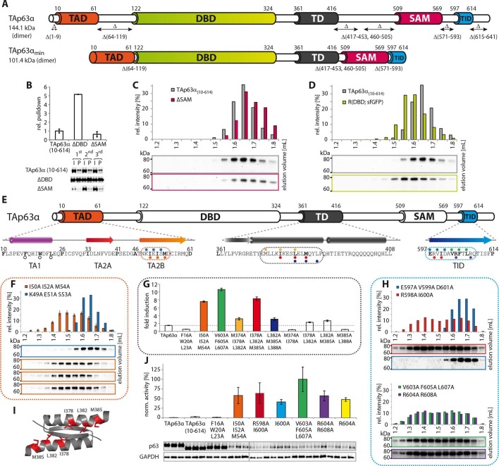
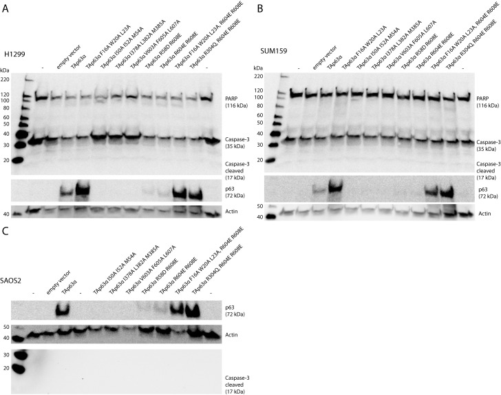
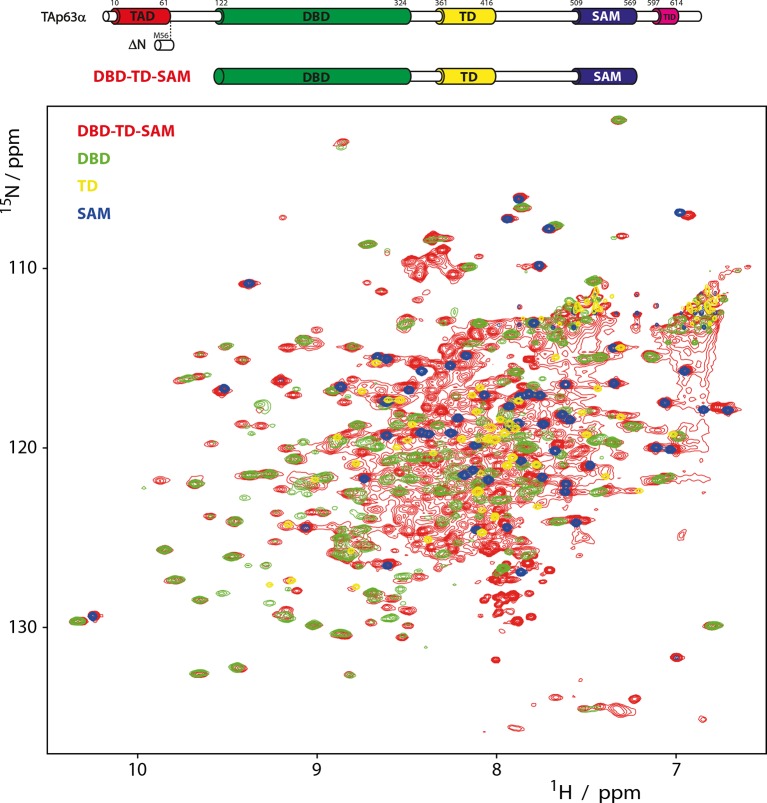
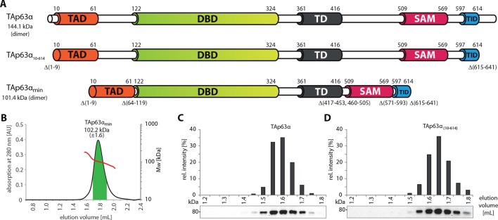
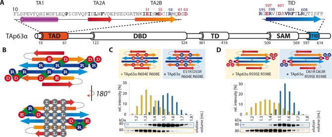
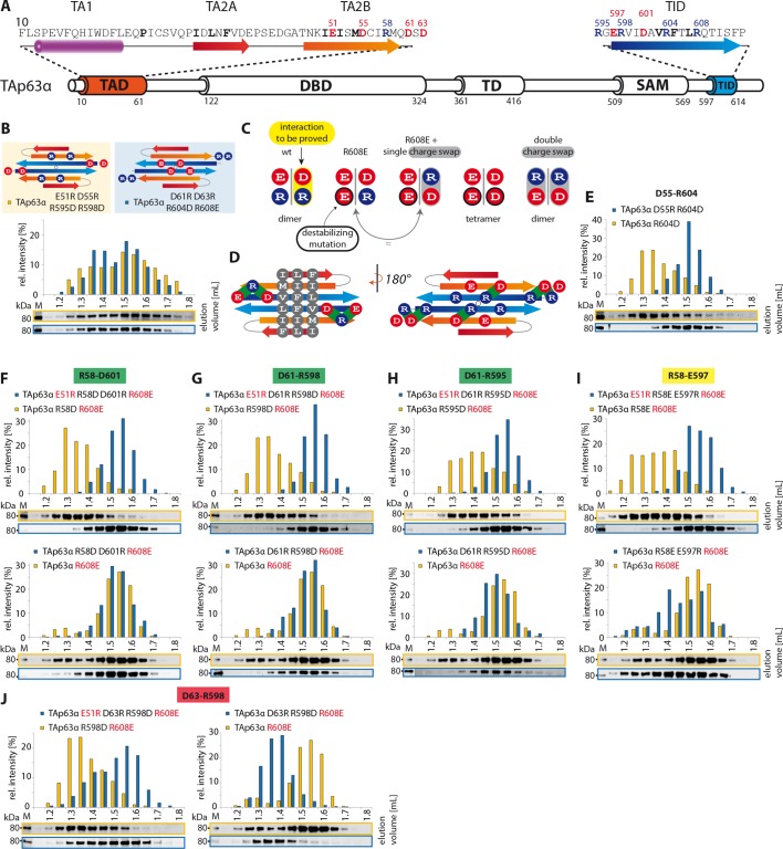
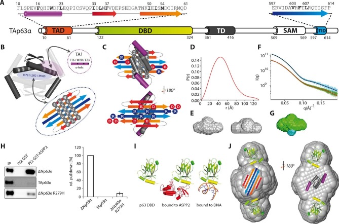
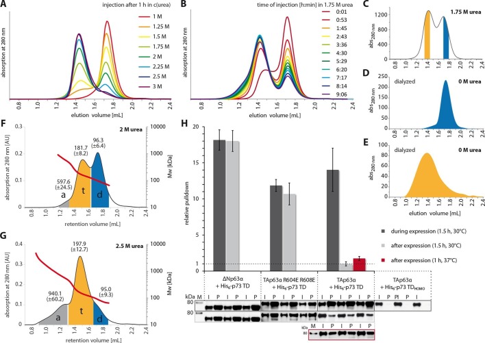
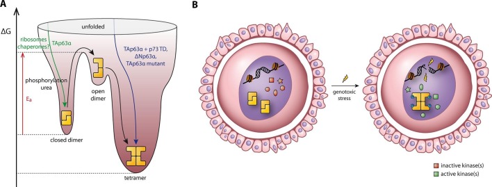
Mammalian oocytes are arrested in the dictyate stage of meiotic prophase I for long periods of time, during which the high concentration of the p53 family member TAp63α sensitizes them to DNA damage-induced apoptosis. TAp63α is kept in an inactive and exclusively dimeric state but undergoes rapid phosphorylation-induced tetramerization and concomitant activation upon detection of DNA damage. Here we show that the TAp63α dimer is a kinetically trapped state. Activation follows a spring-loaded mechanism not requiring further translation of other cellular factors in oocytes and is associated with unfolding of the inhibitory structure that blocks the tetramerization interface. Using a combination of biophysical methods as well as cell and ovary culture experiments we explain how TAp63α is kept inactive in the absence of DNA damage but causes rapid oocyte elimination in response to a few DNA double strand breaks thereby acting as the key quality control factor in maternal reproduction.

摘要

哺乳动物卵母细胞在减数分裂前期I的双线期长时间停滞,在此期间,p53家族成员TAp63α的高浓度使其对DNA损伤诱导的凋亡敏感。TAp63α保持无活性且仅为二聚体状态,但在检测到DNA损伤时会经历快速的磷酸化诱导的四聚化并伴随激活。在这里,我们表明TAp63α二聚体是一种动力学捕获状态。激活遵循一种弹簧加载机制,不需要卵母细胞中其他细胞因子的进一步翻译,并且与阻断四聚化界面的抑制性结构的解折叠有关。通过结合生物物理方法以及细胞和卵巢培养实验,我们解释了TAp63α在没有DNA损伤的情况下如何保持无活性,但在响应少数DNA双链断裂时导致卵母细胞快速消除,从而在母体生殖中作为关键的质量控制因子发挥作用。

https://cdn.ncbi.nlm.nih.gov/pmc/blobs/c588/4876613/f8b999a5af12/elife-13909-resp-fig1.jpg
https://cdn.ncbi.nlm.nih.gov/pmc/blobs/c588/4876613/a9263ff51980/elife-13909-fig1.jpg
https://cdn.ncbi.nlm.nih.gov/pmc/blobs/c588/4876613/bef55907a8c5/elife-13909-fig1-figsupp1.jpg
https://cdn.ncbi.nlm.nih.gov/pmc/blobs/c588/4876613/d39827131600/elife-13909-fig1-figsupp2.jpg
https://cdn.ncbi.nlm.nih.gov/pmc/blobs/c588/4876613/91aa366f8f87/elife-13909-fig1-figsupp3.jpg
https://cdn.ncbi.nlm.nih.gov/pmc/blobs/c588/4876613/54ceef91a2d0/elife-13909-fig1-figsupp4.jpg
https://cdn.ncbi.nlm.nih.gov/pmc/blobs/c588/4876613/27901263a879/elife-13909-fig1-figsupp5.jpg
https://cdn.ncbi.nlm.nih.gov/pmc/blobs/c588/4876613/c561462364d3/elife-13909-fig1-figsupp6.jpg
https://cdn.ncbi.nlm.nih.gov/pmc/blobs/c588/4876613/e3da9e3a1a60/elife-13909-fig1-figsupp7.jpg
https://cdn.ncbi.nlm.nih.gov/pmc/blobs/c588/4876613/b157d9c6fcea/elife-13909-fig1-figsupp8.jpg
https://cdn.ncbi.nlm.nih.gov/pmc/blobs/c588/4876613/a3bd0e3ad617/elife-13909-fig1-figsupp9.jpg
https://cdn.ncbi.nlm.nih.gov/pmc/blobs/c588/4876613/ab4576886843/elife-13909-fig1-figsupp10.jpg
https://cdn.ncbi.nlm.nih.gov/pmc/blobs/c588/4876613/f46eebe94bf8/elife-13909-fig2.jpg
https://cdn.ncbi.nlm.nih.gov/pmc/blobs/c588/4876613/b9fb3cf90574/elife-13909-fig2-figsupp1.jpg
https://cdn.ncbi.nlm.nih.gov/pmc/blobs/c588/4876613/c3a113641bdc/elife-13909-fig3.jpg
https://cdn.ncbi.nlm.nih.gov/pmc/blobs/c588/4876613/81a992764fdc/elife-13909-fig4.jpg
https://cdn.ncbi.nlm.nih.gov/pmc/blobs/c588/4876613/e440cc9ef165/elife-13909-fig4-figsupp1.jpg
https://cdn.ncbi.nlm.nih.gov/pmc/blobs/c588/4876613/24deaf1250e9/elife-13909-fig4-figsupp2.jpg
https://cdn.ncbi.nlm.nih.gov/pmc/blobs/c588/4876613/6d16d61c5dd9/elife-13909-fig5.jpg
https://cdn.ncbi.nlm.nih.gov/pmc/blobs/c588/4876613/f2db32e87a83/elife-13909-fig6.jpg
https://cdn.ncbi.nlm.nih.gov/pmc/blobs/c588/4876613/ae79f3b5f68f/elife-13909-fig6-figsupp1.jpg
https://cdn.ncbi.nlm.nih.gov/pmc/blobs/c588/4876613/35eed1575a4c/elife-13909-fig7.jpg
https://cdn.ncbi.nlm.nih.gov/pmc/blobs/c588/4876613/f8b999a5af12/elife-13909-resp-fig1.jpg

相似文献

1
Quality control in oocytes by p63 is based on a spring-loaded activation mechanism on the molecular and cellular level.p63对卵母细胞的质量控制基于分子和细胞水平上的一种弹簧加载激活机制。
Elife. 2016 Mar 14;5:e13909. doi: 10.7554/eLife.13909.
2
DNA damage in oocytes induces a switch of the quality control factor TAp63α from dimer to tetramer.卵母细胞中的 DNA 损伤会诱导质量控制因子 TAp63α 从二聚体转变为四聚体。
Cell. 2011 Feb 18;144(4):566-76. doi: 10.1016/j.cell.2011.01.013.
3
Defying DNA double-strand break-induced death during prophase I meiosis by temporal TAp63α phosphorylation regulation in developing mouse oocytes.在发育中的小鼠卵母细胞中,通过时间性 TAp63α 磷酸化调节来抵抗减数分裂前期 I 中 DNA 双链断裂诱导的死亡。
Mol Cell Biol. 2014 Apr;34(8):1460-73. doi: 10.1128/MCB.01223-13. Epub 2014 Feb 10.
4
Enhanced pro-apoptosis gene signature following the activation of TAp63α in oocytes upon γ irradiation.γ 辐照激活卵母细胞中的 TAp63α 后,促凋亡基因标志物增强。
Cell Death Dis. 2022 Mar 4;13(3):204. doi: 10.1038/s41419-022-04659-2.
5
p63 null mutation protects mouse oocytes from radio-induced apoptosis.p63基因无效突变可保护小鼠卵母细胞免受辐射诱导的凋亡。
Reproduction. 2008 Jan;135(1):3-12. doi: 10.1530/REP-07-0054.
6
p53 gene family p51(p63)-encoded, secondary transactivator p51B(TAp63alpha) occurs without forming an immunoprecipitable complex with MDM2, but responds to genotoxic stress by accumulation.p53基因家族中由p51(p63)编码的二级反式激活因子p51B(TAp63α)在不与MDM2形成可免疫沉淀复合物的情况下产生,但通过积累对基因毒性应激作出反应。
Exp Cell Res. 2002 Jun 10;276(2):194-200. doi: 10.1006/excr.2002.5535.
7
p63 protects the female germ line during meiotic arrest.p63 在减数分裂停滞期间保护雌性生殖系。
Nature. 2006 Nov 30;444(7119):624-8. doi: 10.1038/nature05337. Epub 2006 Nov 22.
8
Damage limitation.损害控制
Elife. 2016 May 23;5:e17394. doi: 10.7554/eLife.17394.
9
p63 uses a switch-like mechanism to set the threshold for induction of apoptosis.p63 通过一种类似开关的机制来设定细胞凋亡诱导的阈值。
Nat Chem Biol. 2020 Oct;16(10):1078-1086. doi: 10.1038/s41589-020-0600-3. Epub 2020 Jul 27.
10
DNA Damaged Induced Cell Death in Oocytes.DNA 损伤诱导的卵母细胞细胞死亡。
Molecules. 2020 Dec 3;25(23):5714. doi: 10.3390/molecules25235714.

引用本文的文献

1
Differential Transcriptional Activity of ΔNp63β Is Encoded by an Isoform-Specific C-Terminus.ΔNp63β的差异转录活性由一种异构体特异性C末端编码。
Mol Cell Biol. 2025 Jun 23:1-17. doi: 10.1080/10985549.2025.2514529.
2
Alternative splicing in the DBD linker region of p63 modulates binding to DNA and iASPP in vitro.p63的DBD连接区中的可变剪接在体外调节与DNA和iASPP的结合。
Cell Death Dis. 2025 Jan 6;16(1):4. doi: 10.1038/s41419-024-07320-2.
3
Crosstalk between paralogs and isoforms influences p63-dependent regulatory element activity.

本文引用的文献

1
, a program for rapid shape determination in small-angle scattering.用于小角散射中快速形状测定的一个程序。
J Appl Crystallogr. 2009 Apr 1;42(Pt 2):342-346. doi: 10.1107/S0021889809000338. Epub 2009 Jan 24.
2
The Phyre2 web portal for protein modeling, prediction and analysis.用于蛋白质建模、预测和分析的Phyre2网络门户。
Nat Protoc. 2015 Jun;10(6):845-58. doi: 10.1038/nprot.2015.053. Epub 2015 May 7.
3
Gene regulatory mechanisms orchestrated by p63 in epithelial development and related disorders.由p63精心调控的基因调控机制在上皮发育及相关疾病中的作用
旁系同源物和异构体之间的相互作用影响p63依赖性调控元件的活性。
Nucleic Acids Res. 2024 Dec 11;52(22):13812-13831. doi: 10.1093/nar/gkae1143.
4
Oocyte death is triggered by the stabilization of TAp63α dimers in response to cisplatin.卵母细胞死亡是由顺铂诱导的 TAp63α 二聚体的稳定化所触发的。
Cell Death Dis. 2024 Nov 7;15(11):799. doi: 10.1038/s41419-024-07202-7.
5
Advances in the genetic etiology of female infertility.女性不孕症遗传病因学的进展。
J Assist Reprod Genet. 2024 Dec;41(12):3261-3286. doi: 10.1007/s10815-024-03248-w. Epub 2024 Sep 25.
6
Molecular Mechanisms Determining Mammalian Oocyte Quality with the Treatment of Cancer Therapy.决定哺乳动物卵母细胞质量的分子机制及其在癌症治疗中的处理。
Adv Anat Embryol Cell Biol. 2024;238:97-119. doi: 10.1007/978-3-031-55163-5_5.
7
Context dependent activity of p63-bound gene regulatory elements.p63结合基因调控元件的上下文依赖性活性。
bioRxiv. 2024 May 12:2024.05.09.593326. doi: 10.1101/2024.05.09.593326.
8
Fuzzy interactions between the auto-phosphorylated C-terminus and the kinase domain of CK1δ inhibits activation of TAp63α.CK1δ 自身磷酸化的 C 末端与激酶结构域之间的模糊相互作用抑制了 TAp63α 的激活。
Sci Rep. 2023 Sep 30;13(1):16423. doi: 10.1038/s41598-023-43515-x.
9
Heterozygous TP63 pathogenic variants in isolated primary ovarian insufficiency.孤立性原发性卵巢功能不全中杂合性 TP63 致病性变异。
J Assist Reprod Genet. 2023 Sep;40(9):2211-2218. doi: 10.1007/s10815-023-02886-w. Epub 2023 Jul 15.
10
Disease-related p63 DBD mutations impair DNA binding by distinct mechanisms and varying degree.疾病相关的 p63 DBD 突变以不同的机制和不同程度损害 DNA 结合。
Cell Death Dis. 2023 Apr 18;14(4):274. doi: 10.1038/s41419-023-05796-y.
Biochim Biophys Acta. 2015 Jun;1849(6):590-600. doi: 10.1016/j.bbagrm.2015.03.003. Epub 2015 Mar 19.
4
Order and disorder control the functional rearrangement of influenza hemagglutinin.有序和无序控制流感血凝素的功能重排。
Proc Natl Acad Sci U S A. 2014 Aug 19;111(33):12049-54. doi: 10.1073/pnas.1412849111. Epub 2014 Jul 31.
5
Reversal of female infertility by Chk2 ablation reveals the oocyte DNA damage checkpoint pathway.Chk2 缺失可逆转雌性不孕,揭示卵母细胞 DNA 损伤检查点途径。
Science. 2014 Jan 31;343(6170):533-6. doi: 10.1126/science.1247671.
6
Analysis of the oligomeric state and transactivation potential of TAp73α.TAp73α 寡聚状态和反式激活潜力分析。
Cell Death Differ. 2013 Aug;20(8):1008-16. doi: 10.1038/cdd.2013.23. Epub 2013 Mar 29.
7
DNA damage-induced primordial follicle oocyte apoptosis and loss of fertility require TAp63-mediated induction of Puma and Noxa.DNA 损伤诱导原始卵泡卵母细胞凋亡和生育力丧失需要 TAp63 介导的 Puma 和 Noxa 的诱导。
Mol Cell. 2012 Nov 9;48(3):343-52. doi: 10.1016/j.molcel.2012.08.017. Epub 2012 Sep 20.
8
Structural basis of p63α SAM domain mutants involved in AEC syndrome.AEC 综合征相关 p63α SAM 结构域突变体的结构基础。
FEBS J. 2011 Aug;278(15):2680-8. doi: 10.1111/j.1742-4658.2011.08194.x. Epub 2011 Jun 20.
9
Structures of p63 DNA binding domain in complexes with half-site and with spacer-containing full response elements.p63 基因 DNA 结合域与半位点和含间隔子的全长反应元件复合物的结构。
Proc Natl Acad Sci U S A. 2011 Apr 19;108(16):6456-61. doi: 10.1073/pnas.1013657108. Epub 2011 Apr 4.
10
The p53 family: guardians of maternal reproduction.p53 家族:母性生殖的守护者。
Nat Rev Mol Cell Biol. 2011 Apr;12(4):259-65. doi: 10.1038/nrm3086.