Falkai Peter, Steiner Johann, Malchow Berend, Shariati Jawid, Knaus Andreas, Bernstein Hans-Gert, Schneider-Axmann Thomas, Kraus Theo, Hasan Alkomiet, Bogerts Bernhard, Schmitt Andrea
Department of Psychiatry and Psychotherapy, Ludwig Maximilians-University Munich Munich, Germany.
Department of Psychiatry and Psychotherapy, University of Magdeburg Magdeburg, Germany.
Front Cell Neurosci. 2016 Mar 30;10:78. doi: 10.3389/fncel.2016.00078. eCollection 2016.
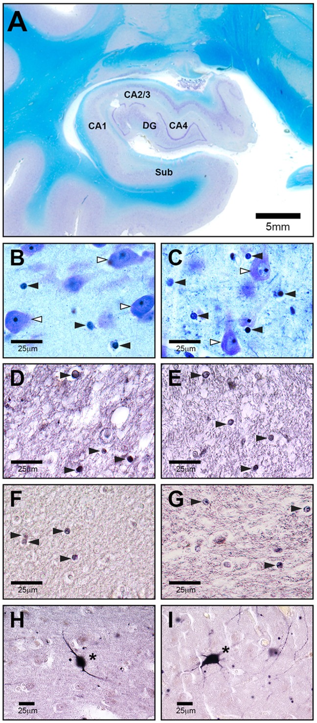
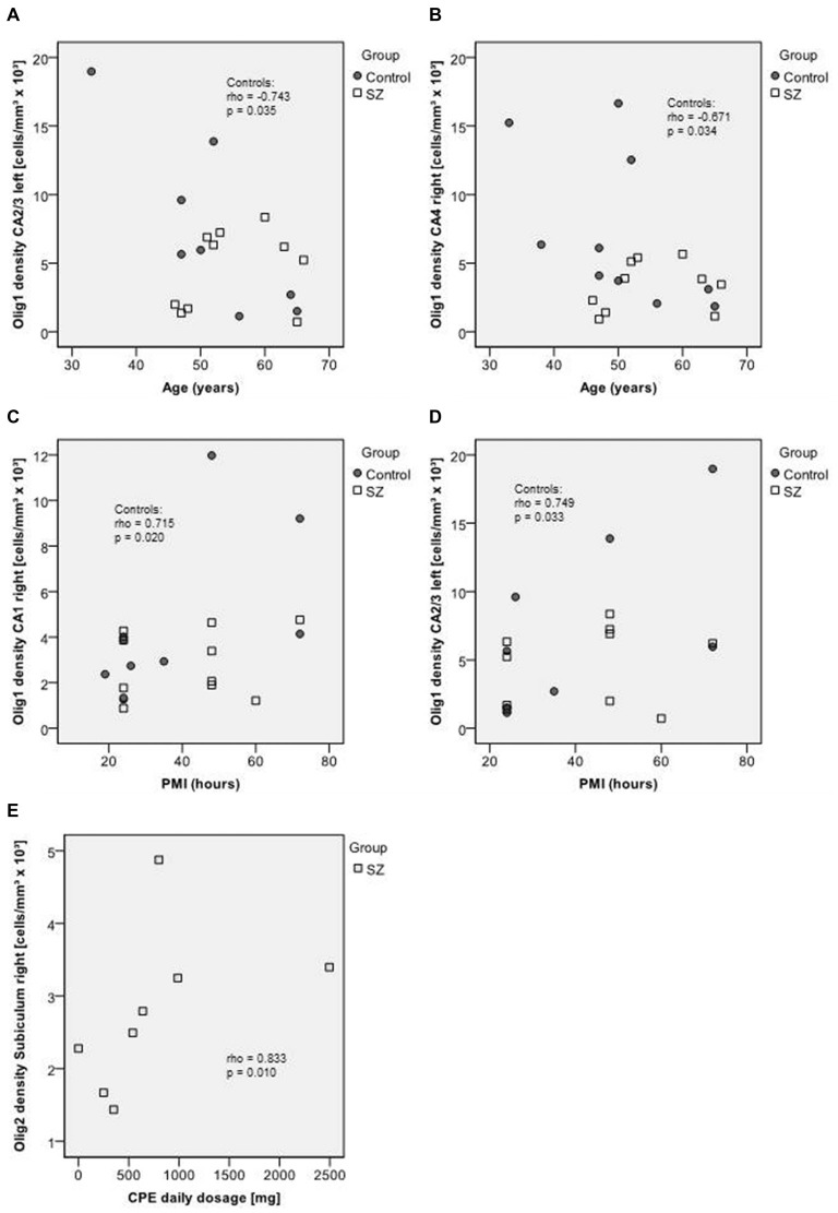
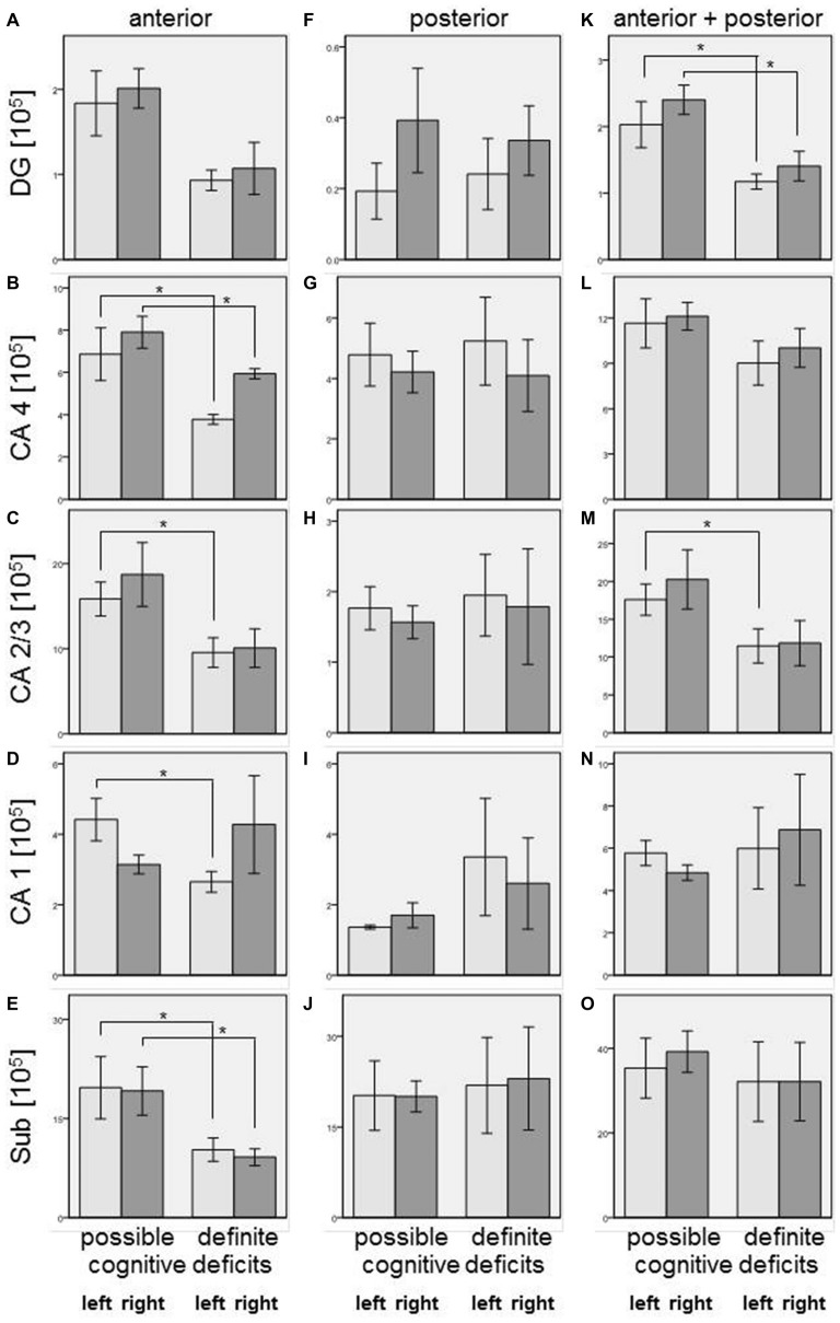
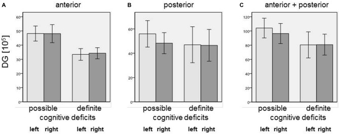
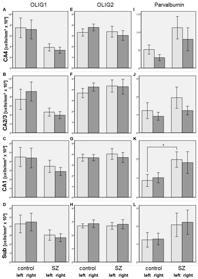
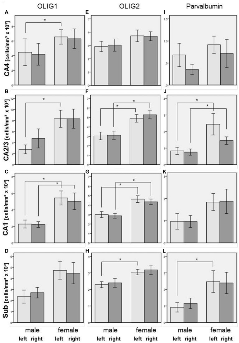
In schizophrenia, previous stereological post-mortem investigations of anterior, posterior, and total hippocampal subfields showed no alterations in total neuron number but did show decreased oligodendrocyte numbers in CA4, an area that corresponds to the polymorph layer of the dentate gyrus (DG). However, these investigations identified oligodendrocytes only on the basis of morphological criteria in Nissl staining and did not assess alterations of interneurons with immunohistochemical markers. Moreover, the association of findings in the posterior hippocampus with cognitive deficits remains unknown. On the basis of the available clinical records, we compared patients with definite and possible cognitive dysfunction; nine patients had evidence in their records of either definite (n = 4) or possible (n = 5) cognitive dysfunction. Additionally, we assessed the density of two oligodendrocyte subpopulations immunostained by the oligodendrocyte transcription factors Olig1 and Olig2 and of interneurons immunolabeled by parvalbumin. We investigated posterior hippocampal subregions in the post-mortem brains of the same schizophrenia patients (SZ; n = 10) and healthy controls (n = 10) we examined in our previously published stereological studies. Our stereological studies found that patients with definite cognitive deficits had decreased total/Nissl-stained oligodendrocyte numbers in the left (p = 0.014) and right (p = 0.050) CA4, left CA2/3 (p = 0.050), left CA1 (p = 0.027), and left (p = 0.050) and right (p = 0.014) subiculum of the anterior part of the hippocampus compared to patients with possible cognitive deficits. In the present study, we found no significant influence of definite cognitive deficits in the posterior part of the hippocampus, whereas in the entire hippocampus SZ with definite cognitive deficits showed decreased oligodendrocyte numbers in the left (p = 0.050) and right (p = 0.050) DG and left CA2/3 (p = 0.050). We did not find significant differences in Olig1-, Olig2-, or parvalbumin-positive cell density between SZ and controls in any of the subregions of the posterior hippocampus. Based on the results from our stereological study we hypothesize that a decreased number of oligodendrocytes in the anterior and entire hippocampus may be involved in cognitive deficits by impairing the connectivity of this structure in schizophrenia. In the posterior hippocampus, we could not replicate previously reported findings of decreased interneurons from the entire hippocampus.
在精神分裂症中,先前对海马前、后亚区及整个海马亚区进行的体视学尸检研究表明,总神经元数量没有改变,但在与齿状回(DG)多形层相对应的CA4区,少突胶质细胞数量确实减少。然而,这些研究仅根据尼氏染色的形态学标准来识别少突胶质细胞,并未用免疫组化标记物评估中间神经元的改变。此外,海马后部的研究结果与认知缺陷之间的关联仍不清楚。根据现有的临床记录,我们比较了有明确和可能存在认知功能障碍的患者;9例患者的记录中有明确(n = 4)或可能(n = 5)认知功能障碍的证据。此外,我们评估了由少突胶质细胞转录因子Olig1和Olig2免疫染色的两个少突胶质细胞亚群以及由小白蛋白免疫标记的中间神经元的密度。我们在先前发表的体视学研究中所检查的同一批精神分裂症患者(SZ;n = 10)和健康对照(n = 10)的尸检大脑中,研究了海马后部亚区。我们的体视学研究发现,与可能存在认知缺陷的患者相比,有明确认知缺陷的患者在海马前部的左侧(p = 0.014)和右侧(p = 0.050)CA4、左侧CA2/3(p = 0.050)、左侧CA1(p = 0.027)以及左侧(p = 0.050)和右侧(p = 0.014)下托中,总/尼氏染色少突胶质细胞数量减少。在本研究中,我们发现海马后部明确的认知缺陷没有显著影响,而在整个海马中,有明确认知缺陷的精神分裂症患者在左侧(p = 0.050)和右侧(p = 0.050)DG以及左侧CA2/3(p = 0.050)的少突胶质细胞数量减少。在海马后部的任何亚区中,我们均未发现精神分裂症患者与对照组之间Olig1、Olig2或小白蛋白阳性细胞密度存在显著差异。基于我们体视学研究的结果,我们推测海马前部和整个海马中少突胶质细胞数量减少可能通过损害精神分裂症中该结构的连接性而参与认知缺陷的形成。在海马后部,我们无法重复先前报道的整个海马中间神经元减少的发现。