Kakite Suguru, Dyvorne Hadrien A, Lee Karen M, Jajamovich Guido H, Knight-Greenfield Ashley, Taouli Bachir
Department of Radiology, Icahn School of Medicine at Mount Sinai, One Gustave Levy Place, New York, NY 10029, USA; Translational and Molecular Imaging Institute, Icahn School of Medicine at Mount Sinai, One Gustave Levy Place, New York, NY 10029, USA.
Translational and Molecular Imaging Institute, Icahn School of Medicine at Mount Sinai, One Gustave Levy Place, New York, NY 10029, USA.
Eur J Radiol Open. 2015 Dec 8;3:1-7. doi: 10.1016/j.ejro.2015.11.002. eCollection 2016.
To correlate intra voxel incoherent motion (IVIM) diffusion parameters of liver parenchyma and hepatocellular carcinoma (HCC) with degree of liver/tumor enhancement and necrosis; and to assess the diagnostic performance of diffusion parameters vs. enhancement ratios (ER) for prediction of complete tumor necrosis.
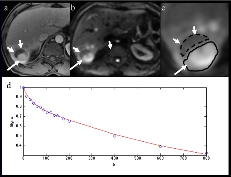
In this IRB approved HIPAA compliant study, we included 46 patients with HCC who underwent IVIM diffusion-weighted (DW) MRI in addition to routine sequences at 3.0 T. True diffusion coefficient (D), pseudo-diffusion coefficient (D*), perfusion fraction (PF) and apparent diffusion coefficient (ADC) were quantified in tumors and liver parenchyma. Tumor ER were calculated using contrast-enhanced imaging, and degree of tumor necrosis was assessed using post-contrast image subtraction. IVIM parameters and ER were compared between HCC and background liver and between necrotic and viable tumor components. ROC analysis for prediction of complete tumor necrosis was performed.
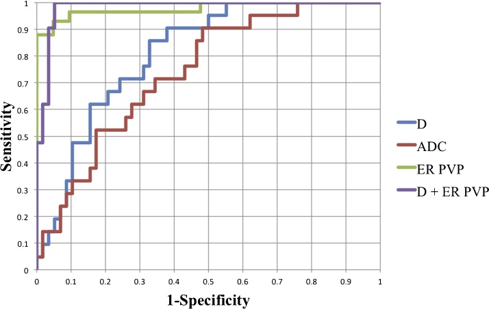
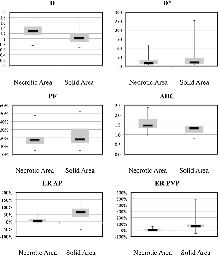
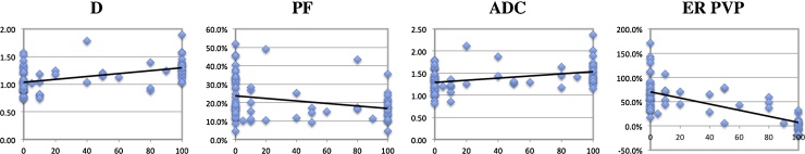
79 HCCs were assessed (mean size 2.5 cm). D, PF and ADC were significantly higher in HCC vs. liver (p < 0.0001). There were weak significant negative/positive correlations between D/PF and ER, and significant correlations between D/PF/ADC and tumor necrosis (for D, r 0.452, p < 0.001). Among diffusion parameters, D had the highest area under the curve (AUC 0.811) for predicting complete tumor necrosis. ER outperformed diffusion parameters for prediction of complete tumor necrosis (AUC > 0.95, p < 0.002).
D has a reasonable diagnostic performance for predicting complete tumor necrosis, however lower than that of contrast-enhanced imaging.
将肝实质和肝细胞癌(HCC)的体素内不相干运动(IVIM)扩散参数与肝脏/肿瘤强化程度及坏死程度相关联;并评估扩散参数与强化率(ER)对预测肿瘤完全坏死的诊断性能。
在这项经机构审查委员会(IRB)批准且符合健康保险流通与责任法案(HIPAA)的研究中,我们纳入了46例HCC患者,这些患者除了在3.0T下进行常规序列检查外,还接受了IVIM扩散加权(DW)磁共振成像(MRI)检查。对肿瘤和肝实质中的真实扩散系数(D)、伪扩散系数(D*)、灌注分数(PF)和表观扩散系数(ADC)进行了定量分析。使用对比增强成像计算肿瘤强化率,并通过对比后图像相减评估肿瘤坏死程度。比较了HCC与肝实质背景之间以及坏死与存活肿瘤成分之间的IVIM参数和强化率。进行了预测肿瘤完全坏死的ROC分析。
共评估了79个HCC(平均大小2.5cm)。HCC中的D、PF和ADC显著高于肝脏(p<0.0001)。D/PF与强化率之间存在弱的显著负/正相关,D/PF/ADC与肿瘤坏死之间存在显著相关(对于D,r=0.452,p<0.001)。在扩散参数中,D在预测肿瘤完全坏死方面的曲线下面积(AUC)最高(AUC=0.811)。在预测肿瘤完全坏死方面,强化率优于扩散参数(AUC>0.95,p<0.002)。
D在预测肿瘤完全坏死方面具有合理的诊断性能,但低于对比增强成像。