Feng Yuan, Kawrakow Iwan, Olsen Jeff, Parikh Parag J, Noel Camille, Wooten Omar, Du Dongsu, Mutic Sasa, Hu Yanle
Soochow University; Washington University School of Medicine; University of Texas at Austin.
J Appl Clin Med Phys. 2016 Mar 8;17(2):441-460. doi: 10.1120/jacmp.v17i2.5820.
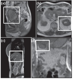
On-board magnetic resonance (MR) image guidance during radiation therapy offers the potential for more accurate treatment delivery. To utilize the real-time image information, a crucial prerequisite is the ability to successfully segment and track regions of interest (ROI). The purpose of this work is to evaluate the performance of different segmentation algorithms using motion images (4 frames per second) acquired using a MR image-guided radiotherapy (MR-IGRT) system. Manual con-tours of the kidney, bladder, duodenum, and a liver tumor by an experienced radiation oncologist were used as the ground truth for performance evaluation. Besides the manual segmentation, images were automatically segmented using thresholding, fuzzy k-means (FKM), k-harmonic means (KHM), and reaction-diffusion level set evolution (RD-LSE) algorithms, as well as the tissue tracking algorithm provided by the ViewRay treatment planning and delivery system (VR-TPDS). The performance of the five algorithms was evaluated quantitatively by comparing with the manual segmentation using the Dice coefficient and target registration error (TRE) measured as the distance between the centroid of the manual ROI and the centroid of the automatically segmented ROI. All methods were able to successfully segment the bladder and the kidney, but only FKM, KHM, and VR-TPDS were able to segment the liver tumor and the duodenum. The performance of the thresholding, FKM, KHM, and RD-LSE algorithms degraded as the local image contrast decreased, whereas the performance of the VP-TPDS method was nearly independent of local image contrast due to the reference registration algorithm. For segmenting high-contrast images (i.e., kidney), the thresholding method provided the best speed (< 1 ms) with a satisfying accuracy (Dice = 0.95). When the image contrast was low, the VR-TPDS method had the best automatic contour. Results suggest an image quality determination procedure before segmentation and a combination of different methods for optimal segmentation with the on-board MR-IGRT system.
放射治疗期间的机载磁共振(MR)图像引导为更精确的治疗提供了可能。为了利用实时图像信息,一个关键前提是能够成功分割并追踪感兴趣区域(ROI)。这项工作的目的是使用磁共振图像引导放射治疗(MR-IGRT)系统采集的运动图像(每秒4帧)评估不同分割算法的性能。由经验丰富的放射肿瘤学家手动勾勒的肾脏、膀胱、十二指肠和肝脏肿瘤轮廓用作性能评估 的标准。除了手动分割外,还使用阈值法、模糊k均值(FKM)、k次谐波均值(KHM)、反应扩散水平集演化(RD-LSE)算法以及ViewRay治疗计划与传输系统(VR-TPDS)提供的组织追踪算法对图像进行自动分割。通过使用Dice系数以及以手动ROI质心与自动分割ROI质心之间的距离衡量的目标配准误差(TRE)与手动分割进行比较,对这五种算法的性能进行定量评估。所有方法都能够成功分割膀胱和肾脏,但只有FKM、KHM和VR-TPDS能够分割肝脏肿瘤和十二指肠。随着局部图像对比度降低,阈值法、FKM、KHM和RD-LSE算法的性能下降,而由于参考配准算法,VP-TPDS方法的性能几乎与局部图像对比度无关。对于分割高对比度图像(即肾脏),阈值法速度最快(<1毫秒)且精度令人满意(Dice = 0.95)。当图像对比度较低时,VR-TPDS方法的自动轮廓效果最佳。结果表明,在分割前需进行图像质量判定,并结合不同方法以在机载MR-IGRT系统中实现最佳分割。