Ren Jianwai, Chen George G, Liu Yi, Su Xianwei, Hu Baoguang, Leung Billy C S, Wang Y, Ho Rocky L K, Yang Shengli, Lu Gang, Lee C G, Lai Paul B S
Department of Surgery, Faculty of Medicine, The Chinese University of Hong Kong; New Territories, Hong Kong, China.
CUHK Shenzhen Research Institute (SZRI), Shenzhen, 518057, China.
PLoS One. 2016 Apr 19;11(4):e0153863. doi: 10.1371/journal.pone.0153863. eCollection 2016.
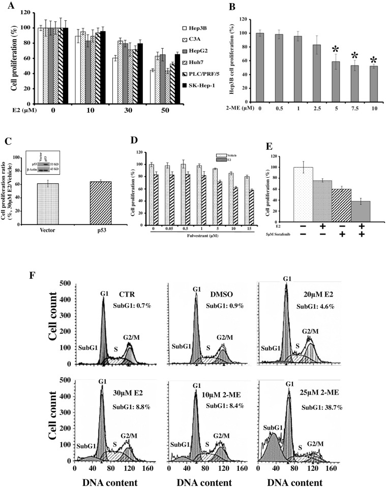
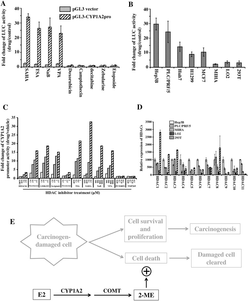
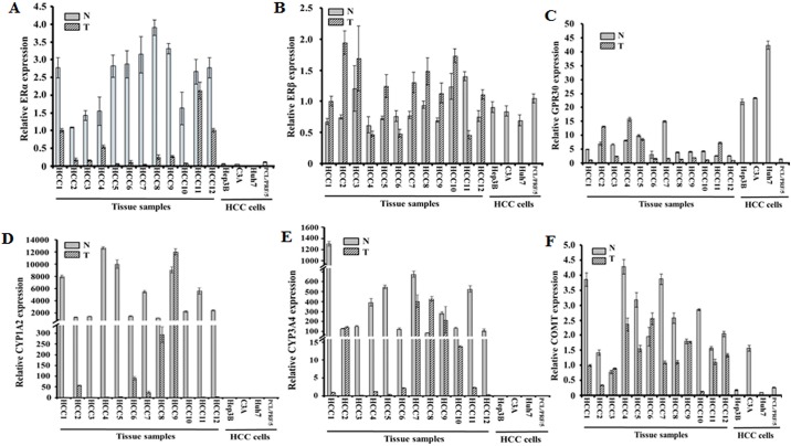
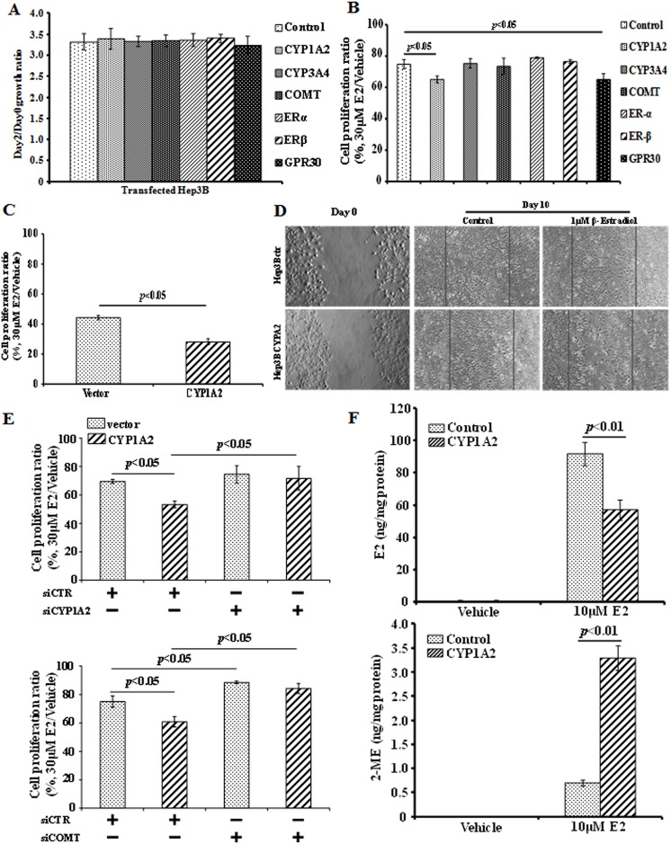
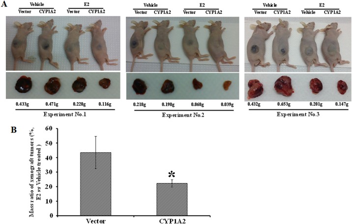
Hepatocellular carcinoma (HCC) occurs more frequently in men than in women. It is commonly agreed that estrogen plays important roles in suppressing HCC development, however, the underlying mechanism remains largely unknown. Since estrogen is mainly metabolized in liver and its metabolites affect cell proliferation, we sought to investigate if the liver-specific cytochrome P450 1A2 (CYP1A2) mediated the inhibitory effect of estrogen on HCC. In this study, the expression of estrogen-metabolizing enzyme CYP1A2 was determined in HCC tissues and cell lines. Cell proliferation and apoptosis were assessed in cells with or without CYP1A2 overexpression. The levels of 17β-estradiol (E2) and its metabolite 2-methoxyestradiol (2-ME) were determined. A xenograft tumor model in mice was established to confirm the findings. It was found that CYP1A2 expression was greatly repressed in HCC. E2 suppressed HCC cell proliferation and xenograft tumor development by inducing apoptosis. The inhibitory effect was significantly enhanced in cells with CYP1A2 overexpression, which effectively conversed E2 to the cytotoxic 2-ME. E2 in combination with sorafenib showed an additive effect on HCC. The anti-HCC effect of E2 was not associated with estrogen receptors ERα and ERβ as well as tumor suppressor P53 but enhanced by the approved anti-HCC drug sorafenib. In addition, HDAC inhibitors greatly induced CYP1A2 promoter activities in cancer cells, especially liver cancer cells, but not in non-tumorigenic cells. Collectively, CYP1A2 metabolizes E2 to generate the potent anti-tumor agent 2-ME in HCC. The reduction of CYP1A2 significantly disrupts this metabolic pathway, contributing the progression and growth of HCC and the gender disparity of this malignancy.
肝细胞癌(HCC)在男性中的发病率高于女性。人们普遍认为雌激素在抑制HCC发展中起重要作用,然而,其潜在机制在很大程度上仍不清楚。由于雌激素主要在肝脏中代谢,其代谢产物影响细胞增殖,我们试图研究肝脏特异性细胞色素P450 1A2(CYP1A2)是否介导雌激素对HCC的抑制作用。在本研究中,检测了HCC组织和细胞系中雌激素代谢酶CYP1A2的表达。评估了过表达或未过表达CYP1A2的细胞的增殖和凋亡情况。测定了17β-雌二醇(E2)及其代谢产物2-甲氧基雌二醇(2-ME)的水平。建立了小鼠异种移植瘤模型以证实研究结果。研究发现,CYP1A2在HCC中表达明显受抑制。E2通过诱导凋亡抑制HCC细胞增殖和异种移植瘤生长。在过表达CYP1A2的细胞中,这种抑制作用显著增强,CYP1A2能有效地将E2转化为具有细胞毒性的2-ME。E2与索拉非尼联合使用对HCC具有相加作用。E2的抗HCC作用与雌激素受体ERα和ERβ以及肿瘤抑制因子P53无关,但被已获批的抗HCC药物索拉非尼增强。此外,组蛋白去乙酰化酶抑制剂能显著诱导癌细胞尤其是肝癌细胞中CYP1A2启动子活性,但对非致瘤细胞无此作用。总的来说,CYP1A2在HCC中将E2代谢生成强效抗肿瘤剂2-ME。CYP1A2的减少显著破坏了这一代谢途径,促进了HCC的进展和生长以及这种恶性肿瘤的性别差异。