Wang Jinju, Guo Runmin, Yang Yi, Jacobs Bradley, Chen Suhong, Iwuchukwu Ifeanyi, Gaines Kenneth J, Chen Yanfang, Simman Richard, Lv Guiyuan, Wu Keng, Bihl Ji C
Department of Pharmacology and Toxicology, Boonshoft School of Medicine, Wright State University, Dayton, OH 45435, USA.
Department of Cardiology, The Affiliated Hospital of Guangdong Medical University, Zhanjiang, Guangdong 524001, China.
Stem Cells Int. 2016;2016:2639728. doi: 10.1155/2016/2639728. Epub 2016 Mar 28.
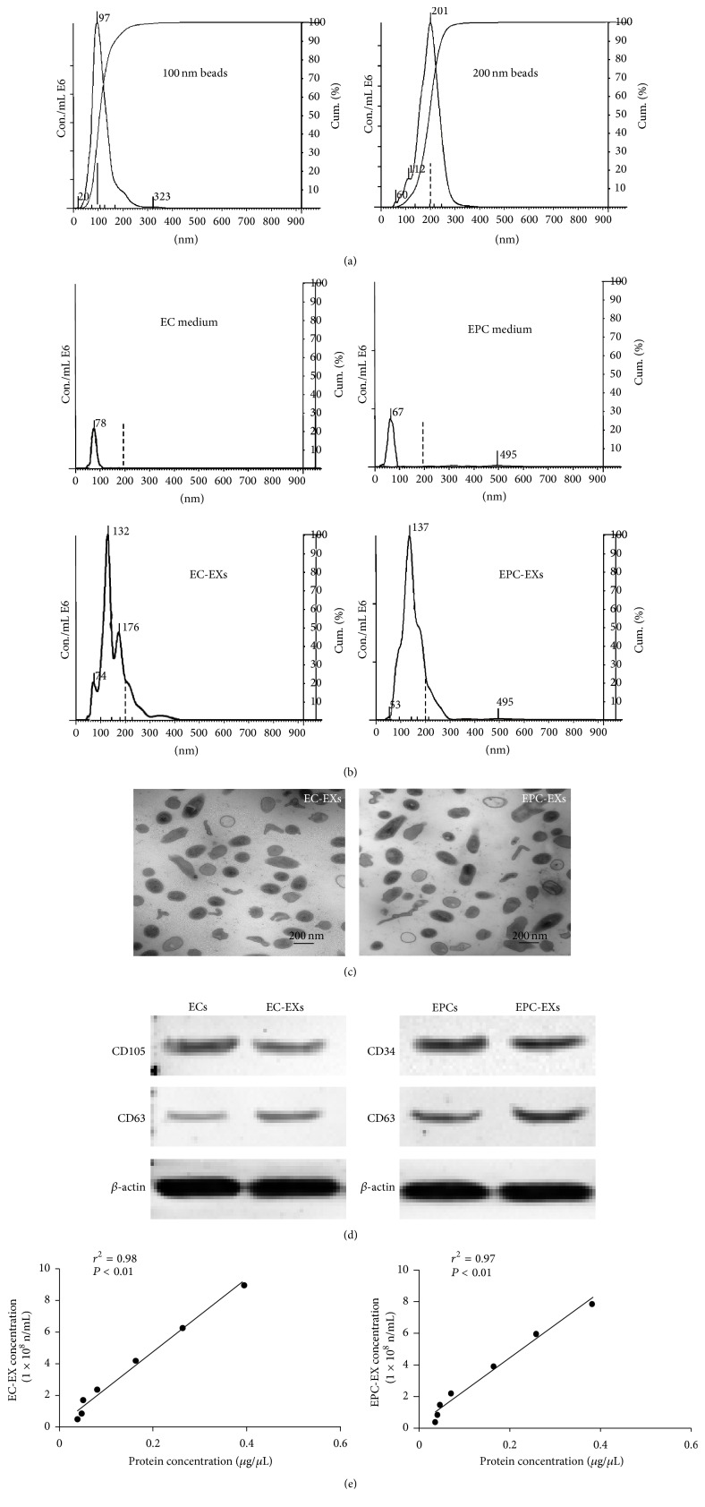
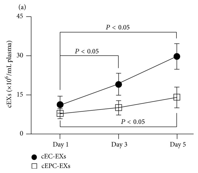
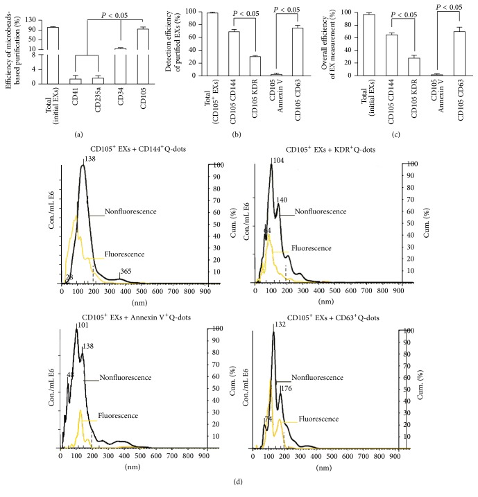
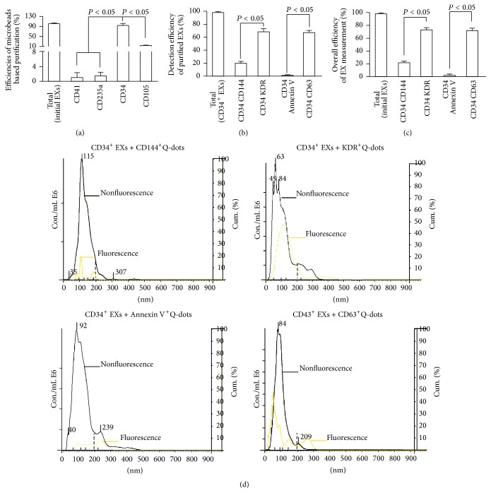
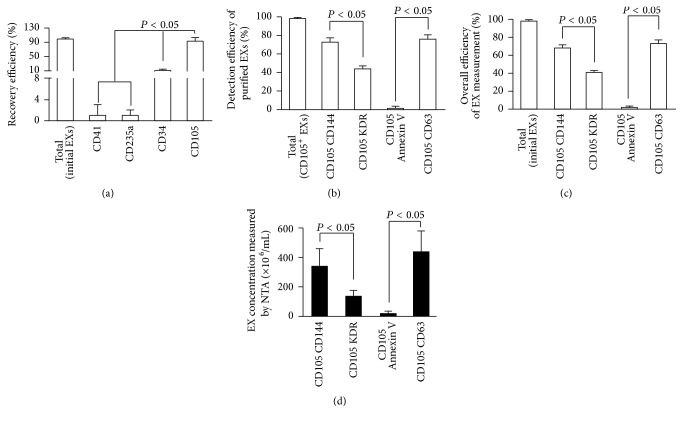
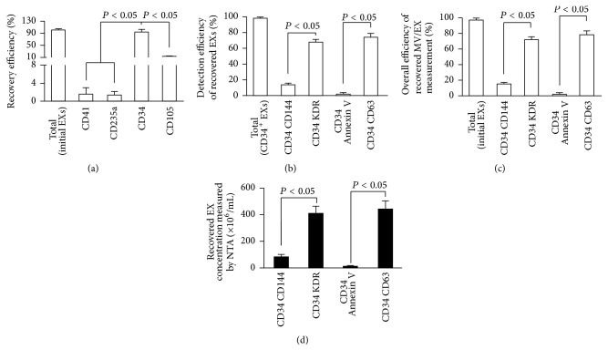
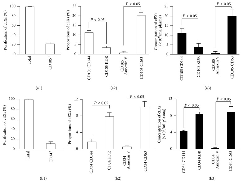
Exosomes (EXs) are cell-derived vesicles that mediate cell-cell communication and could serve as biomarkers. Here we described novel methods for purification and phenotyping of EXs released from endothelial cells (ECs) and endothelial progenitor cells (EPCs) by combining microbeads and fluorescence quantum dots (Q-dots®) techniques. EXs from the culture medium of ECs and EPCs were isolated and detected with cell-specific antibody conjugated microbeads and second antibody conjugated Q-dots by using nanoparticle tracking analysis (NTA) system. The sensitivities of the cell origin markers for ECs (CD105, CD144) and EPCs (CD34, KDR) were evaluated. The sensitivity and specificity were determined by using positive and negative markers for EXs (CD63), platelets (CD41), erythrocytes (CD235a), and microvesicles (Annexin V). Moreover, the methods were further validated in particle-free plasma and patient samples. Results showed that anti-CD105/anti-CD144 and anti-CD34/anti-KDR had the highest sensitivity and specificity for isolating and detecting EC-EXs and EPC-EXs, respectively. The methods had the overall recovery rate of over 70% and were able to detect the dynamical changes of circulating EC-EXs and EPC-EXs in acute ischemic stroke. In conclusion, we have developed sensitive and specific microbeads/Q-dots fluorescence NTA methods for EC-EX and EPC-EX isolation and detection, which will facilitate the functional study and biomarker discovery.
外泌体(EXs)是细胞来源的囊泡,介导细胞间通讯,并可作为生物标志物。在此,我们描述了通过结合微珠和荧光量子点(Q点)技术,从内皮细胞(ECs)和内皮祖细胞(EPCs)中释放的EXs进行纯化和表型分析的新方法。使用纳米颗粒跟踪分析(NTA)系统,用细胞特异性抗体偶联的微珠和二抗偶联的Q点分离和检测ECs和EPCs培养基中的EXs。评估了ECs(CD105、CD144)和EPCs(CD34、KDR)细胞来源标志物的敏感性。通过使用EXs(CD63)、血小板(CD41)、红细胞(CD235a)和微泡(膜联蛋白V)的阳性和阴性标志物来确定敏感性和特异性。此外,该方法在无颗粒血浆和患者样本中得到进一步验证。结果表明,抗CD105/抗CD144和抗CD34/抗KDR分别对分离和检测EC-EXs和EPC-EXs具有最高的敏感性和特异性。该方法的总回收率超过70%,能够检测急性缺血性卒中循环EC-EXs和EPC-EXs的动态变化。总之,我们开发了灵敏且特异的微珠/Q点荧光NTA方法用于EC-EX和EPC-EX的分离和检测,这将有助于功能研究和生物标志物的发现。