Seo Dong-Ho, Lee Kyeong-Seok, Shim Jae-Joon, Yoon Seok-Mann
Department of Neurosurgery, Soonchunhyang University Cheonan Hospital, Cheonan, Korea.
Korean J Neurotrauma. 2014 Apr;10(1):22-5. doi: 10.13004/kjnt.2014.10.1.22. Epub 2014 Apr 30.
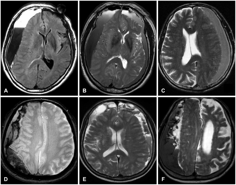
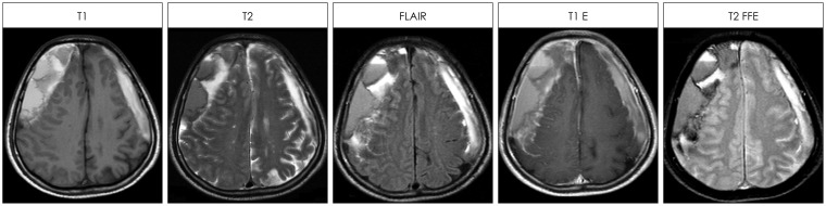
Septa within the hematoma cavity are common, especially in the mixed density chronic subdural hematomas (CSHs). Although CT remains the diagnosis of choice, MRI is superior to detect the membranes in CSHs. We could obtain MRIs in 64 patients with CSH. We examined the value of MRI to understand the history of CSH.
We retrospectively examined the medical records and MRIs of 64 consecutive patients. MRI was selected to find any organic causes of neurologic symptoms. We classified the CSHs into septated or non-septated group, since classification of the septa was frequently obscure.
Septa were identified by MRI in 43 patients (67%). They were more common in the over 70-years-old group. Unknown causes were more common in the septated group, which implies they might suffer from multiple traumas. The signal intensity of the CSH was variable. The methods of treatment were different between two groups. Surgery was more common in the septated group (p=0.021). Surgery was performed in 57 patients (89%). Burr-hole drainage was successful in 55 patients, even in the septated group.
Septa within the hematoma cavity may be related to the multiple episodes of head trauma. Repeated trauma may cause acute bleedings over the CSHs, which is one of the pathogenic mechanisms of hematoma enlargement. MRI could show the history of CSH.
血肿腔内的分隔很常见,尤其是在混合密度的慢性硬膜下血肿(CSH)中。虽然CT仍是首选的诊断方法,但MRI在检测CSH中的包膜方面更具优势。我们对64例CSH患者进行了MRI检查。我们研究了MRI在了解CSH病史方面的价值。
我们回顾性检查了64例连续患者的病历和MRI。选择MRI以查找任何神经系统症状的器质性原因。由于分隔的分类常常不明确,我们将CSH分为有分隔或无分隔组。
43例患者(67%)通过MRI发现有分隔。它们在70岁以上的人群中更为常见。不明原因在有分隔组中更为常见,这意味着他们可能遭受了多次创伤。CSH的信号强度各不相同。两组的治疗方法不同。有分隔组手术更为常见(p=0.021)。57例患者(89%)接受了手术。钻孔引流在55例患者中成功,即使在有分隔组中也是如此。
血肿腔内的分隔可能与头部多次创伤有关。反复创伤可能导致CSH上的急性出血,这是血肿扩大的致病机制之一。MRI可以显示CSH的病史。