Tang Rongxiang, Razi Adeel, Friston Karl J, Tang Yi-Yuan
Department of Psychology, Washington University in St. Louis St. Louis, MO, USA.
The Wellcome Trust Centre for Neuroimaging, University College LondonLondon, UK; Department of Electronic Engineering, NED University of Engineering and TechnologyKarachi, Pakistan.
Front Hum Neurosci. 2016 May 4;10:195. doi: 10.3389/fnhum.2016.00195. eCollection 2016.
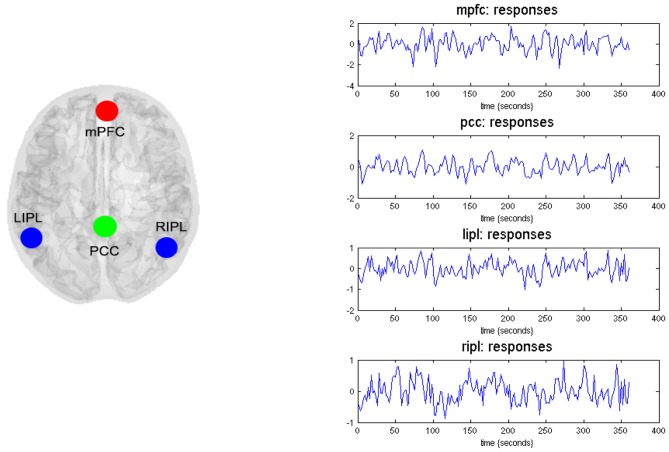
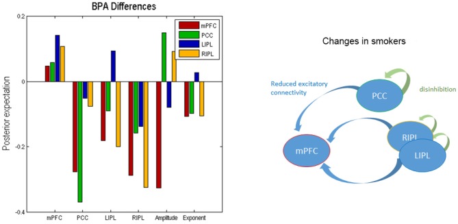
Prefrontal and parietal cortex, including the default mode network (DMN; medial prefrontal cortex (mPFC), and posterior cingulate cortex, PCC), have been implicated in addiction. Nonetheless, it remains unclear which brain regions play a crucial role in smoking addiction and the relationship among these regions. Since functional connectivity only measures correlations, addiction-related changes in effective connectivity (directed information flow) among these distributed brain regions remain largely unknown. Here we applied spectral dynamic causal modeling (spDCM) to resting state fMRI to characterize changes in effective connectivity among core regions in smoking addiction. Compared to nonsmokers, smokers had reduced effective connectivity from PCC to mPFC and from RIPL to mPFC, a higher self-inhibition within PCC and a reduction in the amplitude of endogenous neuronal fluctuations driving the mPFC. These results indicate that spDCM can differentiate the functional architectures between the two groups, and may provide insight into the brain mechanisms underlying smoking addiction. Our results also suggest that future brain-based prevention and intervention in addiction should consider the amelioration of mPFC-PCC-IPL circuits.
前额叶和顶叶皮质,包括默认模式网络(DMN;内侧前额叶皮质(mPFC)和后扣带回皮质,PCC),已被认为与成瘾有关。然而,尚不清楚哪些脑区在吸烟成瘾中起关键作用以及这些区域之间的关系。由于功能连接仅测量相关性,这些分布式脑区之间有效连接(定向信息流)中与成瘾相关的变化在很大程度上仍不为人知。在这里,我们将频谱动态因果模型(spDCM)应用于静息态功能磁共振成像(fMRI),以表征吸烟成瘾核心区域之间有效连接的变化。与不吸烟者相比,吸烟者从PCC到mPFC以及从RIPL到mPFC的有效连接减少,PCC内的自我抑制增强,驱动mPFC的内源性神经元波动幅度降低。这些结果表明,spDCM可以区分两组之间的功能结构,并可能为吸烟成瘾的脑机制提供见解。我们的结果还表明,未来基于脑的成瘾预防和干预应考虑改善mPFC-PCC-IPL回路。