Watson H L, Dybvig K, Blalock D K, Cassell G H
Department of Microbiology, University of Alabama at Birmingham 35294.
Infect Immun. 1989 Jun;57(6):1684-90. doi: 10.1128/iai.57.6.1684-1690.1989.
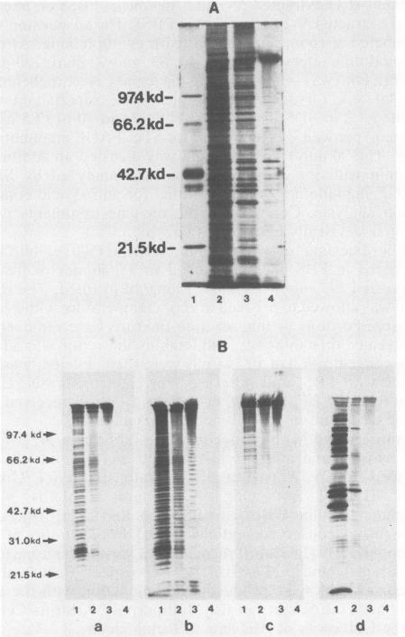
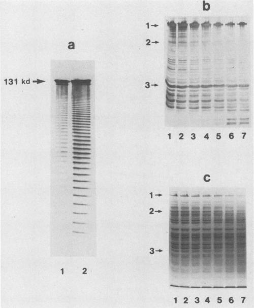
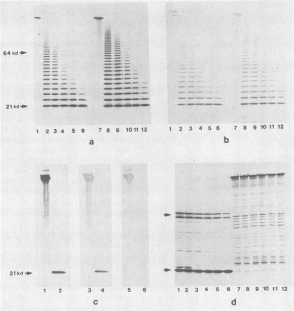
It was previously shown that multiple structural variants of the V-1 antigen (variable antigen 1) of Mycoplasma pulmonis could be found within a single strain. This antigen is unusual in that it produces a ladder pattern after sodium dodecyl sulfate (SDS)-polyacrylamide gel electrophoresis. The present study showed that some variants of V-1 could be extracted into the aqueous phase of a phenol-H2O system. Analysis with anti-V-1 monoclonal antibodies showed that the phenol-H2O-extracted V-1 had a regular spacing of 3.1 kilodaltons (kDa) between bands and trypsinization of this extracted V-1 resulted in the gradual symmetrical collapse (2.9-kDa increments) of the ladder into a single band, suggesting the presence of multiple identical subunits within the V-1 structure. The upper band from the phenol-H2O-extracted V-1 was isolated and analyzed by SDS-polyacrylamide gel electrophoresis immunoblotting, resulting in the regeneration of the original ladder pattern with 3.1-kDa spacing between bands. When V-1 was boiled for increasing times in the presence of SDS, the staining intensity of the upper band decreased with the concurrent appearance of additional lower-molecular-weight bands. Finally, by using whole cells, it was found that the lower-molecular-weight species of the ladder pattern selectively partitioned into the hydrophobic phase of a Triton X-114 phase partitioning system, and the higher-molecular-weight bands were found in the aqueous phase. These data indicate that the V-1 bands are composed of subunits which may aggregate via hydrophobic interactions and that these aggregates at least partially dissociate when exposed to harsh denaturing conditions, resulting in the characteristic ladder pattern of V-1.
先前的研究表明,在肺炎支原体的单个菌株中可以发现V-1抗原(可变抗原1)的多种结构变体。这种抗原不同寻常之处在于,在十二烷基硫酸钠(SDS)-聚丙烯酰胺凝胶电泳后会产生阶梯状条带模式。本研究表明,一些V-1变体可以被提取到苯酚-H₂O系统的水相中。用抗V-1单克隆抗体分析表明,经苯酚-H₂O提取的V-1条带之间的间距规则,为3.1千道尔顿(kDa),对这种提取的V-1进行胰蛋白酶消化会导致阶梯状条带逐渐对称塌陷(以2.9-kDa的增量)为单一的条带,这表明V-1结构中存在多个相同的亚基。分离出经苯酚-H₂O提取的V-1的上条带,并通过SDS-聚丙烯酰胺凝胶电泳免疫印迹进行分析,结果重新出现了条带间距为3.1-kDa的原始阶梯状条带模式。当V-1在SDS存在下煮沸不同时间时,上条带的染色强度降低,同时出现额外的低分子量条带。最后,使用完整细胞发现,阶梯状条带模式中较低分子量的物种选择性地分配到Triton X-114相分配系统的疏水相中,而较高分子量的条带存在于水相中。这些数据表明,V-1条带由亚基组成,这些亚基可能通过疏水相互作用聚集,并且当暴露于苛刻的变性条件时,这些聚集体至少部分解离,从而产生V-1特有的阶梯状条带模式。