Lu Yi, Liang Hongmin, Han Dan, Mo Yin, Li Zongfang, Cheng Yuqi, Xu Xiufeng, Shen Zonglin, Tan Chunyan, Zhao Wei, Zhu Yun, Sun Xuejin
Department of Medical Imaging, The First Affiliated Hospital, Kunming Medical University, Kunming, Yunnan, China.
Department of Psychiatry, The First Affiliated Hospital, Kunming Medical University, Kunming, Yunnan, China.
Neuroimage Clin. 2016 May 14;11:658-666. doi: 10.1016/j.nicl.2016.04.008. eCollection 2016.
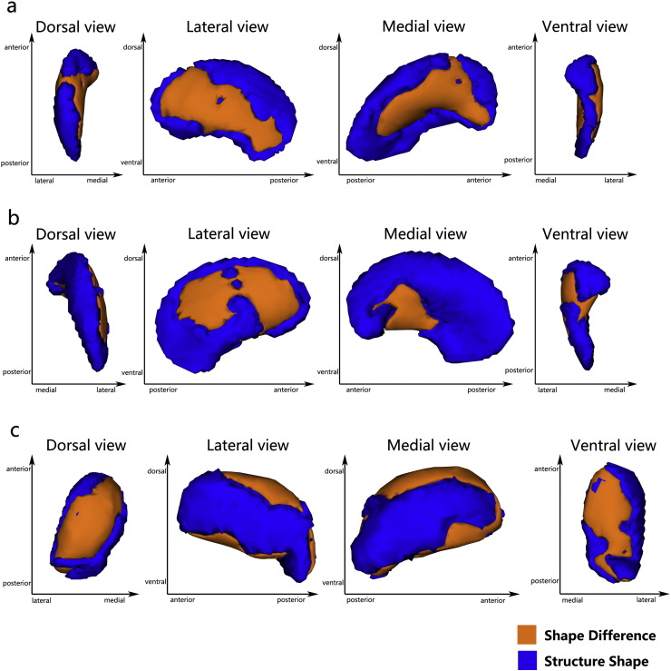
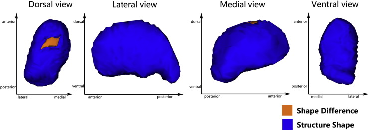
Previous MRI studies confirmed abnormalities in the limbic-cortical-striatal-pallidal-thalamic (LCSPT) network or limbic-cortico-striatal-thalamic-cortical (LCSTC) circuits in patients with major depressive disorder (MDD), but few studies have investigated the subcortical structural abnormalities. Therefore, we sought to determine whether focal subcortical grey matter (GM) changes might be present in MDD at an early stage. We recruited 30 first episode, untreated patients with major depressive disorder (MDD) and 26 healthy control subjects. Voxel-based morphometry was used to evaluate cortical grey matter changes, and automated volumetric and shape analyses were used to assess volume and shape changes of the subcortical GM structures, respectively. In addition, probabilistic tractography methods were used to demonstrate the relationship between the subcortical and the cortical GM. Compared to healthy controls, MDD patients had significant volume reductions in the bilateral putamen and left thalamus (FWE-corrected, p < 0.05). Meanwhile, the vertex-based shape analysis showed regionally contracted areas on the dorsolateral and ventromedial aspects of the bilateral putamen, and on the dorsal and ventral aspects of left thalamus in MDD patients (FWE-corrected, p < 0.05). Additionally, a negative correlation was found between local atrophy in the dorsal aspects of the left thalamus and clinical variables representing severity. Furthermore, probabilistic tractography demonstrated that the area of shape deformation of the bilateral putamen and left thalamus have connections with the frontal and temporal lobes, which were found to be related to major depression. Our results suggested that structural abnormalities in the putamen and thalamus might be present in the early stages of MDD, which support the role of subcortical structure in the pathophysiology of MDD. Meanwhile, the present study showed that these subcortical structural abnormalities might be the potential trait markers of MDD.
先前的磁共振成像(MRI)研究证实,重度抑郁症(MDD)患者的边缘-皮质-纹状体-苍白球-丘脑(LCSPT)网络或边缘-皮质-纹状体-丘脑-皮质(LCSTC)回路存在异常,但很少有研究调查皮质下结构异常。因此,我们试图确定在MDD早期是否可能存在局灶性皮质下灰质(GM)变化。我们招募了30例首次发作、未接受治疗的重度抑郁症(MDD)患者和26名健康对照者。基于体素的形态测量法用于评估皮质灰质变化,自动体积分析和形状分析分别用于评估皮质下GM结构的体积和形状变化。此外,概率纤维束成像方法用于证明皮质下GM与皮质GM之间的关系。与健康对照相比,MDD患者双侧壳核和左侧丘脑体积显著减小(FWE校正,p<0.05)。同时,基于顶点的形状分析显示,MDD患者双侧壳核的背外侧和腹内侧以及左侧丘脑的背侧和腹侧区域有局部收缩(FWE校正,p<0.05)。此外,发现左侧丘脑背侧局部萎缩与代表严重程度的临床变量之间存在负相关。此外,概率纤维束成像表明,双侧壳核和左侧丘脑的形状变形区域与额叶和颞叶有连接,而这些区域被发现与重度抑郁症有关。我们的结果表明,壳核和丘脑的结构异常可能在MDD早期就存在,这支持了皮质下结构在MDD病理生理学中的作用。同时,本研究表明这些皮质下结构异常可能是MDD的潜在特征标志物。