Bahri Maede, Farrahi Hassan, Bahri Maryam, Mahdavinataj Hami, Batouli Seyed Amir Hossein
Department of Neuroscience and Addiction Studies, School of Advanced Technologies in Medicine, Tehran University of Medical Sciences, Tehran, Iran.
Department of Psychiatry, Kavosh Cognitive Behavior Sciences and Addiction Research Center, School of Medicine, Guilan University of Medical Sciences, Rasht, Iran.
Medicine (Baltimore). 2025 Apr 11;104(15):e42000. doi: 10.1097/MD.0000000000042000.
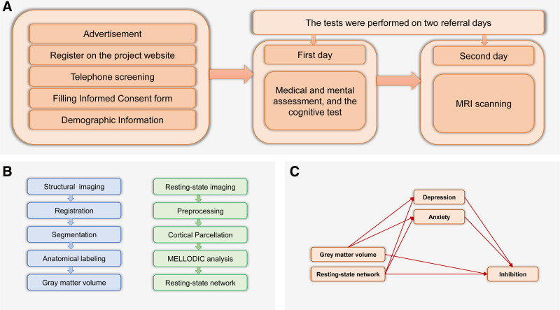
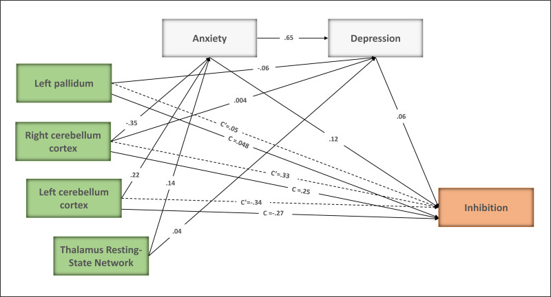
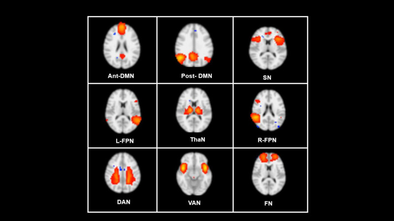
Evidence shows that depression and anxiety symptoms are associated with reduced cognitive inhibition. Nevertheless, the neural substrates responsible for the effects of depression and anxiety symptoms on cognitive inhibition are yet to be determined. This cross-sectional study adhered to the strengthening the reporting of observational studies in epidemiology (STROBE) checklist. Data from 242 participants from the Iranian brain imaging database were used in this study. To address the neural substrates of depression and anxiety responsible for inhibition, voxel-based morphometry (VBM) analysis and resting-state functional magnetic resonance imaging (RS-fMRI) were used. The depression anxiety stress scale was used to evaluate symptoms of depression and anxiety, and the Stroop test was used for cognitive inhibition. The behavioral results demonstrated that inhibition was significantly negatively correlated with depression and anxiety. The VBM results showed that depression was negatively correlated with gray matter (GM) volume in the left pallidum and the right cerebellum cortex. Additionally, anxiety negatively correlated with GM volume in the left and right cerebellum cortex. RS-fMRI results showed that the thalamus network was positively correlated with depression and anxiety. more importantly, mediation analysis revealed that the right cerebellum cortex and thalamic resting-state network through depression and anxiety had a total indirect effect on inhibition. Clarifying the neural substrates responsible for how depression and anxiety symptoms affect cognitive inhibition could have important implications for interventions aimed at supporting individuals' cognitive health.
证据表明,抑郁和焦虑症状与认知抑制能力下降有关。然而,抑郁和焦虑症状对认知抑制产生影响的神经基础尚未确定。这项横断面研究遵循了流行病学观察性研究报告强化(STROBE)清单。本研究使用了来自伊朗脑成像数据库的242名参与者的数据。为了探究抑郁和焦虑影响抑制作用的神经基础,采用了基于体素的形态学测量(VBM)分析和静息态功能磁共振成像(RS-fMRI)。使用抑郁焦虑压力量表评估抑郁和焦虑症状,并使用斯特鲁普测试来评估认知抑制能力。行为学结果表明,抑制能力与抑郁和焦虑显著负相关。VBM结果显示,抑郁与左侧苍白球和右侧小脑皮质的灰质(GM)体积呈负相关。此外,焦虑与左右小脑皮质的GM体积均呈负相关。RS-fMRI结果显示,丘脑网络与抑郁和焦虑呈正相关。更重要的是,中介分析表明,右侧小脑皮质和丘脑静息态网络通过抑郁和焦虑对抑制能力产生了总的间接影响。阐明抑郁和焦虑症状如何影响认知抑制的神经基础,可能对旨在支持个体认知健康的干预措施具有重要意义。