Myhre Adam P, Jarosz Todd J, Hunter John C, Richardson Michael L
Radiol Case Rep. 2015 Nov 6;1(1):21-3. doi: 10.2484/rcr.v1i1.9. eCollection 2006.
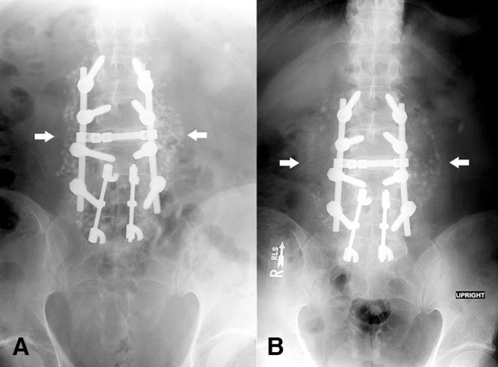
We describe a case of a spinal fusion site infection, which was first suggested on plain radiographs due to interval displacement and partial dissolution of the bone graft material. These radiographic findings occurred 4 weeks before the infection became clinically evident. Cultures taken during eventual surgical debridement grew Aspergillus fumigatus. This case emphasizes the importance of noting changes in bone graft material in addition to the routine evaluation of alignment and hardware in patients who have undergone posterior spinal fusion.
我们描述了一例脊柱融合部位感染病例,最初在X线平片上发现,原因是骨移植材料出现间隔移位和部分溶解。这些影像学表现出现在感染临床症状明显前4周。最终手术清创时采集的培养物培养出烟曲霉。该病例强调了在对接受后路脊柱融合术的患者进行对线和内固定装置常规评估之外,注意骨移植材料变化的重要性。