Lin Chiu-Mei, Chang Hang, Wang Bao-Wei, Shyu Kou-Gi
Department of Emergency Medicine, Shin Kong Wu Ho-Su Memorial Hospital, Taipei, Taiwan.
Faculty of Medicine, School of Medicine, Fu Jen Catholic University, Taipei, Taiwan.
J Cell Mol Med. 2016 Nov;20(11):2045-2055. doi: 10.1111/jcmm.12895. Epub 2016 Jun 16.
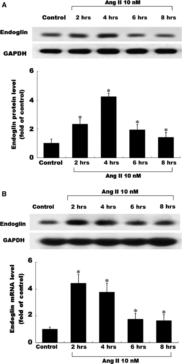
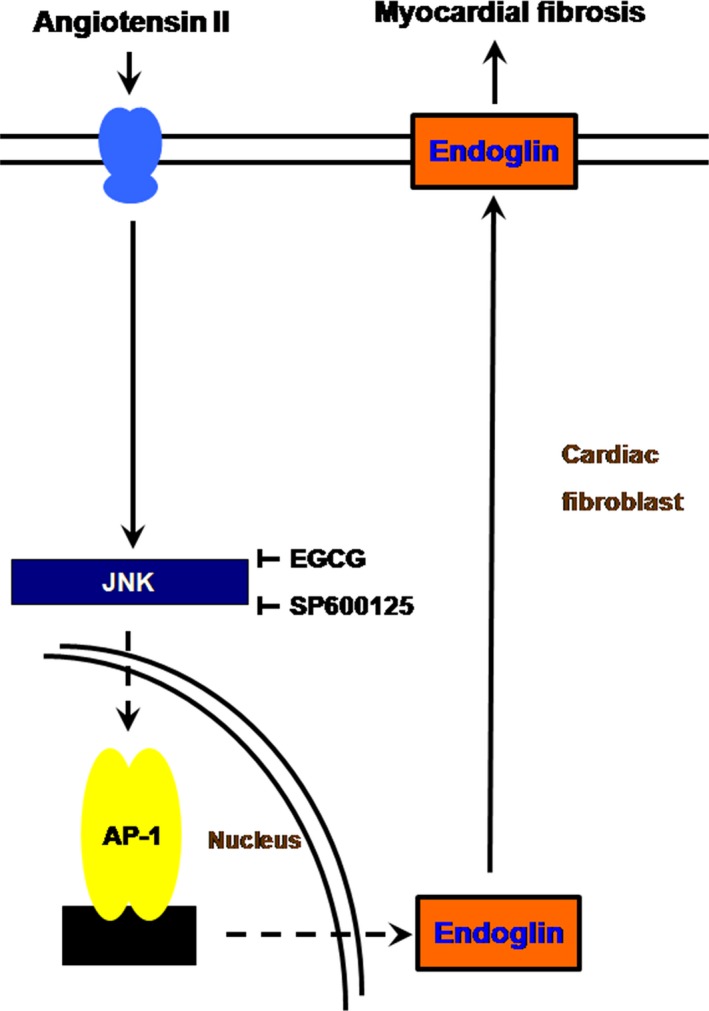
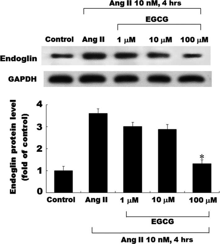
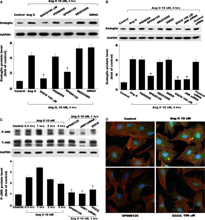
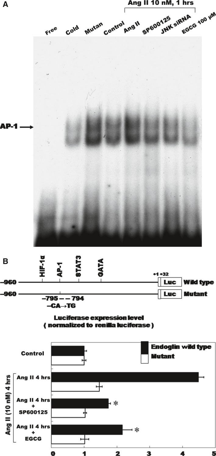
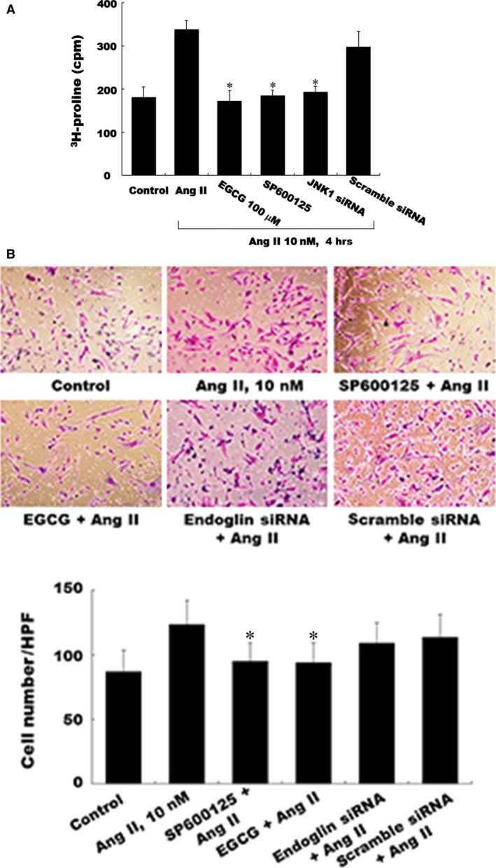
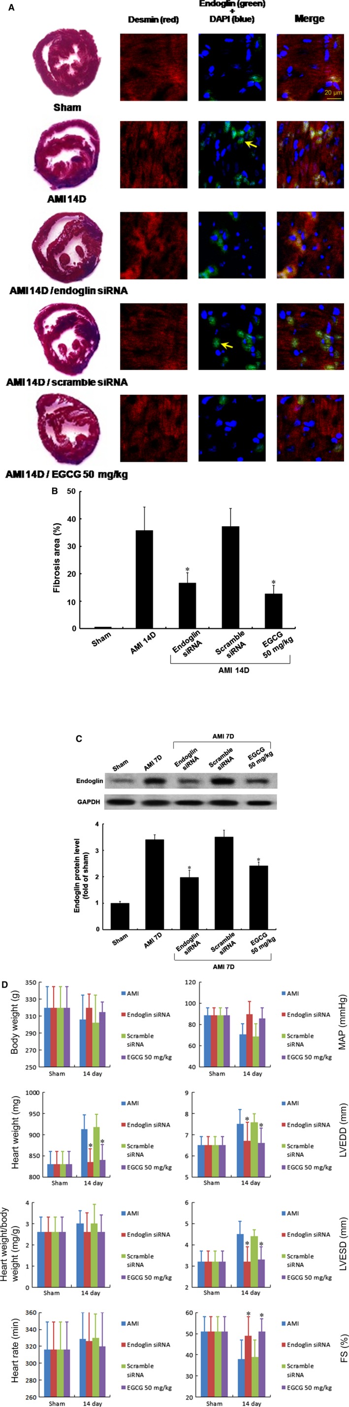
Epigallocatechin-3-O-gallate (EGCG), derived from green tea, has been studied extensively because of its diverse physiological and pharmacological properties. This study evaluates the protective effect of EGCG on angiotensin II (Ang II)-induced endoglin expression in vitro and in vivo. Cardiac fibroblasts (CFs) from the thoracic aorta of adult Wistar rats were cultured and induced with Ang II. Western blotting, Northern blotting, real-time PCR and promoter activity assay were performed. Ang II increased endoglin expression significantly as compared with control cells. The specific extracellular signal-regulated kinase inhibitor SP600125 (JNK inhibitor), EGCG (100 μM) and c-Jun N-terminal kinase (JNK) siRNA attenuated endoglin proteins following Ang II induction. In addition, pre-treated Ang II-induced endoglin with EGCG diminished the binding activity of AP-1 by electrophoretic mobility shift assay. Moreover, the luciferase assay results revealed that EGCG suppressed the endoglin promoter activity in Ang II-induced CFs by AP-1 binding. Finally, EGCG and the JNK inhibitor (SP600125) were found to have attenuated endoglin expression significantly in Ang II-induced CFs, as determined through confocal microscopy. Following in vivo acute myocardial infarction (AMI)-related myocardial fibrosis study, as well as immunohistochemical and confocal analyses, after treatment with endoglin siRNA and EGCG (50 mg/kg), the area of myocardial fibrosis reduced by 53.4% and 64.5% and attenuated the left ventricular end-diastolic and systolic dimensions, and friction shortening in hemodynamic monitor. In conclusion, epigallocatechin-3-O-gallate (EGCG) attenuated the endoglin expression and myocardial fibrosis by anti-inflammatory effect in vitro and in vivo, the novel suppressive effect was mediated through JNK/AP-1 pathway.
表没食子儿茶素-3-没食子酸酯(EGCG)源自绿茶,因其具有多种生理和药理特性而受到广泛研究。本研究评估了EGCG在体外和体内对血管紧张素II(Ang II)诱导的内皮糖蛋白表达的保护作用。培养成年Wistar大鼠胸主动脉的心脏成纤维细胞(CFs)并用Ang II进行诱导。进行了蛋白质免疫印迹法、Northern印迹法、实时聚合酶链反应和启动子活性测定。与对照细胞相比,Ang II显著增加了内皮糖蛋白的表达。特异性细胞外信号调节激酶抑制剂SP600125(JNK抑制剂)、EGCG(100μM)和c-Jun氨基末端激酶(JNK)小干扰RNA在Ang II诱导后减弱了内皮糖蛋白的表达。此外,用EGCG预处理Ang II诱导的内皮糖蛋白后,通过电泳迁移率变动分析减少了AP-1的结合活性。此外,荧光素酶测定结果显示,EGCG通过AP-1结合抑制了Ang II诱导的CFs中的内皮糖蛋白启动子活性。最后,通过共聚焦显微镜检查发现,EGCG和JNK抑制剂(SP600125)在Ang II诱导的CFs中显著减弱了内皮糖蛋白的表达。在体内急性心肌梗死(AMI)相关心肌纤维化研究以及免疫组织化学和共聚焦分析后,在用内皮糖蛋白小干扰RNA和EGCG(50mg/kg)治疗后,心肌纤维化面积分别减少了53.4%和64.5%,并减弱了左心室舒张末期和收缩期内径以及血流动力学监测中的缩短分数。总之,表没食子儿茶素-3-没食子酸酯(EGCG)在体外和体内通过抗炎作用减弱了内皮糖蛋白表达和心肌纤维化作用,这种新的抑制作用是通过JNK/AP-1途径介导的。