• 文献检索
  • 文档翻译
  • 深度研究
  • 学术资讯
  • Suppr Zotero 插件Zotero 插件
  • 邀请有礼
  • 套餐&价格
  • 历史记录
应用&插件
Suppr Zotero 插件Zotero 插件浏览器插件Mac 客户端Windows 客户端微信小程序
定价
高级版会员购买积分包购买API积分包
服务
文献检索文档翻译深度研究API 文档MCP 服务
关于我们
关于 Suppr公司介绍联系我们用户协议隐私条款
关注我们

Suppr 超能文献

核心技术专利:CN118964589B侵权必究
粤ICP备2023148730 号-1Suppr @ 2026

文献检索

告别复杂PubMed语法,用中文像聊天一样搜索,搜遍4000万医学文献。AI智能推荐,让科研检索更轻松。

立即免费搜索

文件翻译

保留排版,准确专业,支持PDF/Word/PPT等文件格式,支持 12+语言互译。

免费翻译文档

深度研究

AI帮你快速写综述,25分钟生成高质量综述,智能提取关键信息,辅助科研写作。

立即免费体验

黑色素瘤切除术后诊断出的细胞性蓝痣:诊断难题

Cellular Blue Nevus Diagnosed following Excision of Melanoma: A Challenge in Diagnosis.

作者信息

Jonjić Nives, Dekanić Andrea, Glavan Nedeljka, Prpić-Massari Larisa, Grahovac Blaženka

机构信息

Department of Pathology, Rijeka University School of Medicine, Braće Branchetta 20, 51000 Rijeka, Croatia.

Department of Pediatric Surgery, Rijeka University Hospital Center, Rijeka, Croatia.

出版信息

Case Rep Pathol. 2016;2016:8107671. doi: 10.1155/2016/8107671. Epub 2016 May 26.

DOI:10.1155/2016/8107671
PMID:27313934
原文链接:https://pmc.ncbi.nlm.nih.gov/articles/PMC4899595/
Abstract

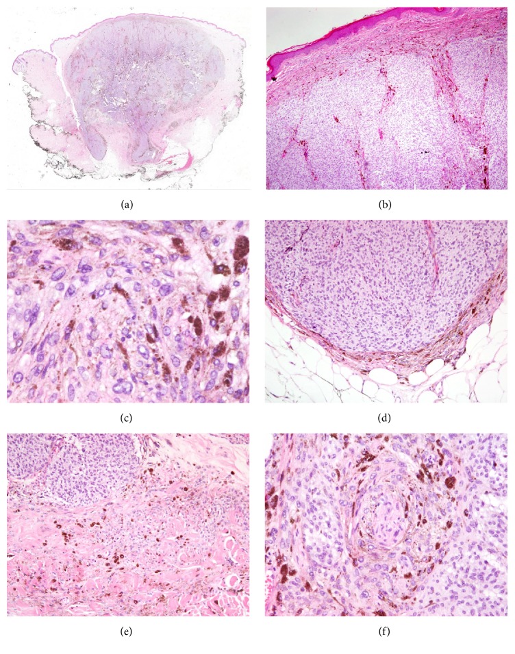
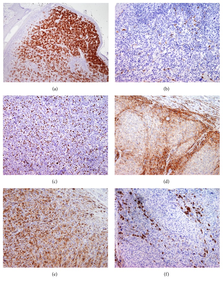
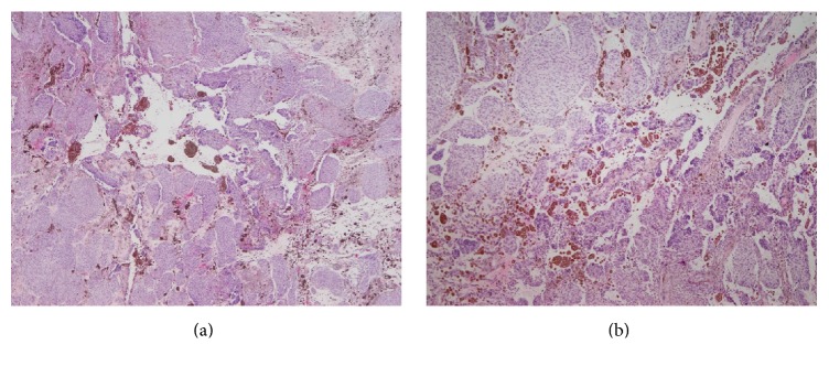
A case of a 41-year-old woman with a history of nodular melanoma (NM), associated with an indurated dome-shaped blue-black nodule with a diameter of 1.2 cm in the gluteal region, is presented. Clinical diagnosis of the lesion, present from birth, was blue nevus. Recently, the nodule has been showing a mild enlargement and thus complete resection was performed. Histological analysis revealed a pigmented lesion with an expansive pattern of extension into the dermis and the subcutaneous adipose tissue. The lesion displayed an alveolar pattern as well as a pigmented dendritic cell pattern. The histology was consistent with cellular blue nevus (CBN); however, the history of NM which was excised one year earlier, as well as the clinical information about the slow growing lesion, included a differential diagnosis of CBN, borderline melanocytic tumor, and malignant blue nevus. Additional immunohistochemical (HMB-45, p16, and Ki-67) and molecular (BRAF V600E mutation) analyses were performed on both lesions: the CBN-like and the previously excised NM. Along with lesion history and histological analyses, p16 staining and BRAF were useful diagnostic tools for confirming the benign nature of CBN in this case.

摘要

本文报告一例41岁女性患者,有结节性黑色素瘤(NM)病史,其臀区有一个直径1.2厘米的硬结性圆顶状蓝黑色结节。该病变自出生就存在,临床诊断为蓝痣。最近,该结节有轻度增大,因此进行了完整切除。组织学分析显示,有一个色素沉着病变,呈向真皮和皮下脂肪组织浸润性生长模式。该病变表现为腺泡状模式以及色素性树突状细胞模式。组织学与细胞性蓝痣(CBN)相符;然而,一年前切除的NM病史以及该病变生长缓慢的临床信息,包括了对CBN、交界性黑素细胞肿瘤和恶性蓝痣的鉴别诊断。对两个病变,即类似CBN的病变和先前切除的NM,进行了额外的免疫组化(HMB-45、p16和Ki-67)及分子(BRAF V600E突变)分析。结合病变病史和组织学分析,p16染色和BRAF在本病例中是确认CBN良性性质的有用诊断工具。

https://cdn.ncbi.nlm.nih.gov/pmc/blobs/4b71/4899595/c2922c83f92e/CRIPA2016-8107671.003.jpg
https://cdn.ncbi.nlm.nih.gov/pmc/blobs/4b71/4899595/bc86115dbec7/CRIPA2016-8107671.001.jpg
https://cdn.ncbi.nlm.nih.gov/pmc/blobs/4b71/4899595/213dee7c975f/CRIPA2016-8107671.002.jpg
https://cdn.ncbi.nlm.nih.gov/pmc/blobs/4b71/4899595/c2922c83f92e/CRIPA2016-8107671.003.jpg
https://cdn.ncbi.nlm.nih.gov/pmc/blobs/4b71/4899595/bc86115dbec7/CRIPA2016-8107671.001.jpg
https://cdn.ncbi.nlm.nih.gov/pmc/blobs/4b71/4899595/213dee7c975f/CRIPA2016-8107671.002.jpg
https://cdn.ncbi.nlm.nih.gov/pmc/blobs/4b71/4899595/c2922c83f92e/CRIPA2016-8107671.003.jpg

相似文献

1
Cellular Blue Nevus Diagnosed following Excision of Melanoma: A Challenge in Diagnosis.黑色素瘤切除术后诊断出的细胞性蓝痣:诊断难题
Case Rep Pathol. 2016;2016:8107671. doi: 10.1155/2016/8107671. Epub 2016 May 26.
2
Pilonidal sinus associated with cellular blue nevus. A previously unrecognized association.
J Cutan Pathol. 2007 Dec;34(12):942-5. doi: 10.1111/j.1600-0560.2007.00753.x.
3
Role of In Vivo Reflectance Confocal Microscopy in the Analysis of Melanocytic Lesions.体内反射共聚焦显微镜在黑素细胞性病变分析中的作用
Acta Dermatovenerol Croat. 2018 Apr;26(1):64-67.
4
Meyerson Phenomenon as a Component of Melanoma in situ.迈耶森现象作为原位黑色素瘤的一个组成部分。
Acta Dermatovenerol Croat. 2016 Apr;24(1):81-2.
5
Large plaque-type blue nevus with subcutaneous cellular nodules.伴有皮下细胞结节的大片状蓝痣
Am J Surg Pathol. 2000 Jan;24(1):92-9. doi: 10.1097/00000478-200001000-00011.
6
Cellular blue nevus (CBN) lymph node metastases of the neck with no primary skin lesion: a case report and review of literature.颈部细胞蓝色神经瘤(CBN)淋巴结转移而无原发性皮肤病变:病例报告及文献复习。
J Craniomaxillofac Surg. 2010 Dec;38(8):601-4. doi: 10.1016/j.jcms.2010.01.008. Epub 2010 Mar 12.
7
Atypical cellular blue nevi (cellular blue nevi with atypical features): lack of consensus for diagnosis and distinction from cellular blue nevi and malignant melanoma ("malignant blue nevus").非典型细胞性蓝痣(具有非典型特征的细胞性蓝痣):在诊断以及与细胞性蓝痣和恶性黑色素瘤(“恶性蓝痣”)的鉴别方面缺乏共识。
Am J Surg Pathol. 2008 Jan;32(1):36-44. doi: 10.1097/PAS.0b013e3181573aaf.
8
p16 Expression Is Lost in Severely Atypical Cellular Blue Nevi and Melanoma Compared to Conventional, Mildly, and Moderately Atypical Cellular Blue Nevi.与传统、轻度和中度非典型细胞性蓝痣相比,p16表达在重度非典型细胞性蓝痣和黑色素瘤中缺失。
ISRN Dermatol. 2014 Jan 22;2014:348417. doi: 10.1155/2014/348417. eCollection 2014.
9
Giant cellular blue nevus of the anterior chest wall mimicking metastatic melanoma to the breast: a case report.前胸壁巨大细胞性蓝色痣酷似乳腺癌转移黑色素瘤:一例报告
J Surg Oncol. 2000 Aug;74(4):278-81. doi: 10.1002/1096-9098(200008)74:4<278::aid-jso7>3.0.co;2-d.
10
A cellular blue nevus with pigmented epithelioid melanocytoma-like pattern on the ipsilateral upper arm associated with a congenital plaque-type blue nevus on the hand.一名患者在同侧上臂出现具有色素性上皮样黑素细胞瘤样模式的细胞性蓝痣,同时手部伴有先天性斑块型蓝痣。
J Cutan Pathol. 2019 May;46(5):383-388. doi: 10.1111/cup.13435. Epub 2019 Mar 28.

引用本文的文献

1
Benign Nevi Mimicking Melanoma: A Diagnostic Dilemma.酷似黑色素瘤的良性痣:诊断难题
Cureus. 2024 Nov 30;16(11):e74821. doi: 10.7759/cureus.74821. eCollection 2024 Nov.
2
Nevus Blue as a Sporadic Finding in a Patient with a Blue Toe?蓝色痣作为蓝趾患者的偶发表现?
Open Access Maced J Med Sci. 2018 May 15;6(5):855-858. doi: 10.3889/oamjms.2018.228. eCollection 2018 May 20.
3
Histologic mimics of malignant melanoma.恶性黑色素瘤的组织学模仿物。

本文引用的文献

1
Molecular alterations in malignant blue nevi and related blue lesions.恶性蓝痣及相关蓝色病变中的分子改变。
Virchows Arch. 2015 Dec;467(6):723-732. doi: 10.1007/s00428-015-1851-3. Epub 2015 Sep 24.
2
p16 Expression Is Lost in Severely Atypical Cellular Blue Nevi and Melanoma Compared to Conventional, Mildly, and Moderately Atypical Cellular Blue Nevi.与传统、轻度和中度非典型细胞性蓝痣相比,p16表达在重度非典型细胞性蓝痣和黑色素瘤中缺失。
ISRN Dermatol. 2014 Jan 22;2014:348417. doi: 10.1155/2014/348417. eCollection 2014.
3
NRAS and BRAF mutations in melanoma-associated nevi and uninvolved nevi.
Singapore Med J. 2018 Nov;59(11):602-607. doi: 10.11622/smedj.2018041. Epub 2018 May 18.
黑色素瘤相关痣和未受累痣中的NRAS 和 BRAF 突变。
PLoS One. 2013 Jul 8;8(7):e69639. doi: 10.1371/journal.pone.0069639. Print 2013.
4
Blue nevi and variants: an update.蓝色痣和变异:更新。
Arch Pathol Lab Med. 2011 Mar;135(3):327-36. doi: 10.5858/2009-0733-RA.1.
5
Polyclonality of BRAF mutations in acquired melanocytic nevi.获得性黑素细胞痣中BRAF突变的多克隆性。
J Natl Cancer Inst. 2009 Oct 21;101(20):1423-7. doi: 10.1093/jnci/djp309. Epub 2009 Sep 14.
6
So-called "malignant blue nevus": a clinicopathologic study of 23 patients.所谓“恶性蓝痣”:23例患者的临床病理研究
Cancer. 2009 Jul 1;115(13):2949-55. doi: 10.1002/cncr.24319.
7
Allelotyping, microsatellite instability, and BRAF mutation analyses in common and atypical melanocytic nevi and primary cutaneous melanomas.常见及非典型黑素细胞痣和原发性皮肤黑色素瘤的基因分型、微卫星不稳定性及BRAF突变分析
Am J Dermatopathol. 2009 Jun;31(4):354-63. doi: 10.1097/DAD.0b013e318185d205.
8
Immunohistochemical characteristics of melanoma.黑色素瘤的免疫组织化学特征
J Cutan Pathol. 2008 May;35(5):433-44. doi: 10.1111/j.1600-0560.2007.00891.x.
9
Atypical cellular blue nevi (cellular blue nevi with atypical features): lack of consensus for diagnosis and distinction from cellular blue nevi and malignant melanoma ("malignant blue nevus").非典型细胞性蓝痣(具有非典型特征的细胞性蓝痣):在诊断以及与细胞性蓝痣和恶性黑色素瘤(“恶性蓝痣”)的鉴别方面缺乏共识。
Am J Surg Pathol. 2008 Jan;32(1):36-44. doi: 10.1097/PAS.0b013e3181573aaf.
10
Effects of G-protein mutations on skin color.G蛋白突变对肤色的影响。
Nat Genet. 2004 Sep;36(9):961-8. doi: 10.1038/ng1412. Epub 2004 Aug 22.