Uberti Francesca, Morsanuto Vera, Lattuada Debora, Colciaghi Barbara, Cochis Andrea, Bulfoni Alessandro, Colombo Paola, Bolis Giorgio, Molinari Claudio
Physiology Laboratory, Department of Translational Medicine, UPO - University of Eastern Piedmont, Via Solaroli 17, Novara, 28100, Italy.
Department of Obstetrics and Gynecology, Fondazione IRCCS Cà Granda, Ospedale Maggiore Policlinico, Milan, 20122, Italy.
J Ovarian Res. 2016 Jun 17;9(1):34. doi: 10.1186/s13048-016-0243-x.
Recently, vitamin D3 (1alpha, 25-dihydroxyvitamin D) has shown its capability to take part in many extraskeletal functions and its serum levels have been related to patient survival rate and malignancy of many types of neoplasms, including ovarian cancers. Catalytic iron is a free circulating form of iron that is able to generate reactive oxygen species and consequently to promote a number of cellular and tissutal dysfunctions including tumorigenesis. In fertile women an important source of catalytic iron is derived from retrograde menstruation. Epithelial secretory cells from fimbriae of fallopian tubes are greatly exposed to catalytic iron derived from menstrual reflux and so represent the site of origin for most serous ovarian cancers. The aim of this study was to assess whether vitamin D3 can play a role in counteracting catalytic iron-induced oxidative stress in cells from fimbriae of fallopian tubes.
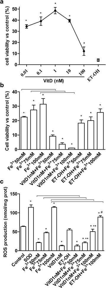
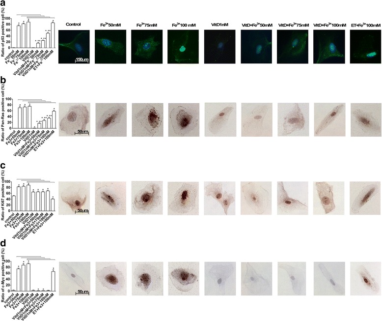
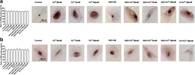
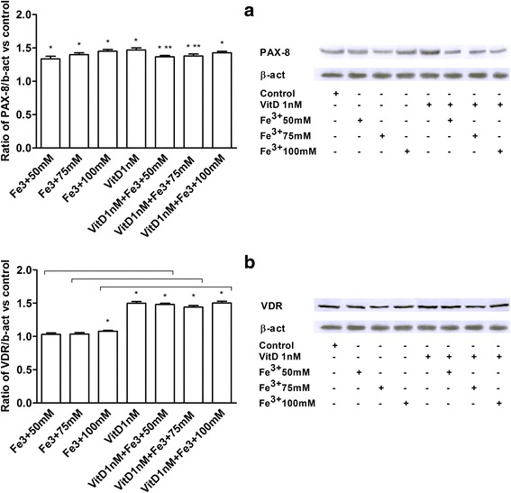
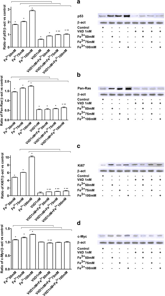
The cells, isolated from women undergoing isteroannessiectomy, were treated with catalytic iron 50-75-100 mM and vitamin D3 at a concentration ranging from 0.01 to 10 nM to study cell viability, radical oxygen species production, p53, pan-Ras, Ki67 and c-Myc protein expressions through Western Blot, and immunocytochemistry or immunofluorescence analysis.
The pre-treatment with vitamin D3 1 nM showed its beneficial effects that consists in a significant decrease in ROS production. In addition a novel finding is represented by the demonstration that pre-treatment with vitamin D3 is also able to significantly counteract tumoral biomarkers activation, such as p53, pan-Ras, Ki67 and c-Myc, and consequently the catalytic iron-induced cellular injury.
This study demonstrates for the first time that vitamin D3 plays an important role in preventing catalytic iron-dependent oxidative stress in cultured fimbrial cells. These results support the hypothesis that vitamin D3 could counteract carcinogenic changes induced by catalytic iron.
最近,维生素D3(1α,25-二羟基维生素D)已显示出参与许多骨骼外功能的能力,其血清水平与包括卵巢癌在内的多种肿瘤的患者生存率和恶性程度相关。催化铁是铁的一种游离循环形式,能够产生活性氧,从而促进包括肿瘤发生在内的许多细胞和组织功能障碍。在育龄妇女中,催化铁的一个重要来源是逆行月经。输卵管伞端的上皮分泌细胞大量暴露于月经反流产生的催化铁中,因此是大多数浆液性卵巢癌的起源部位。本研究的目的是评估维生素D3是否能在对抗催化铁诱导的输卵管伞端细胞氧化应激中发挥作用。
从接受子宫切除术的女性中分离出细胞,用50 - 75 - 100 mM的催化铁和浓度范围为0.01至10 nM的维生素D3进行处理,通过蛋白质免疫印迹、免疫细胞化学或免疫荧光分析来研究细胞活力、活性氧产生、p53、泛Ras、Ki67和c-Myc蛋白表达。
1 nM维生素D3预处理显示出其有益作用,包括活性氧产生显著减少。此外,一个新发现是,维生素D3预处理还能够显著对抗肿瘤生物标志物的激活,如p53、泛Ras、Ki67和c-Myc,从而对抗催化铁诱导的细胞损伤。
本研究首次证明维生素D3在预防培养的伞端细胞中依赖催化铁的氧化应激方面发挥重要作用。这些结果支持维生素D3可以对抗催化铁诱导的致癌变化这一假说。