Blixt Frank W, Johansson Sara Ellinor, Johnson Leif, Haanes Kristian Agmund, Warfvinge Karin, Edvinsson Lars
Department of Clinical Sciences, Division of Experimental Vascular Research, Lund University, Lund, Sweden.
Department of Clinical Experimental Research, Glostrup Research Institute, Rigshospitalet, Glostrup, Denmark.
PLoS One. 2016 Jun 20;11(6):e0157669. doi: 10.1371/journal.pone.0157669. eCollection 2016.
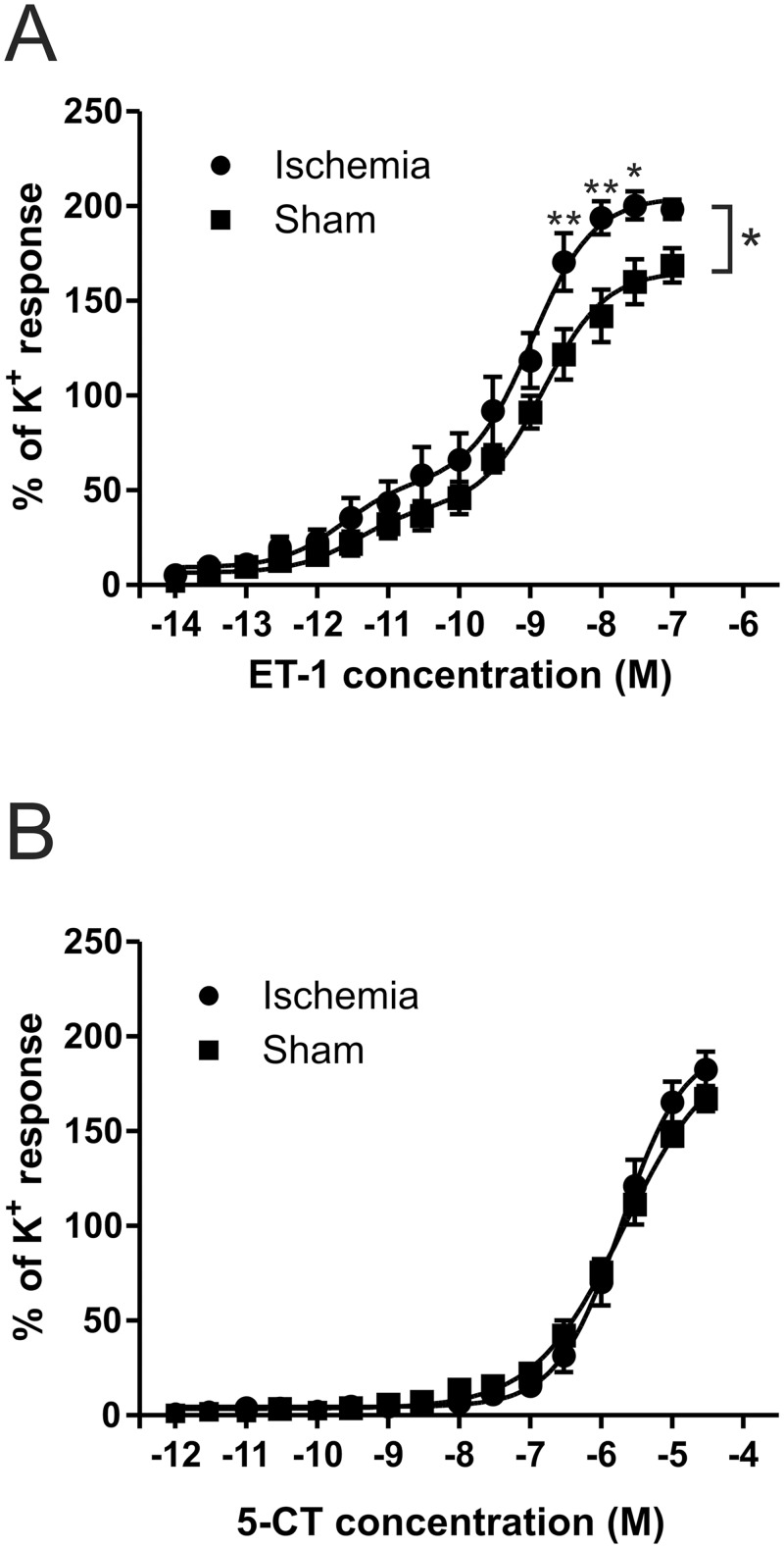
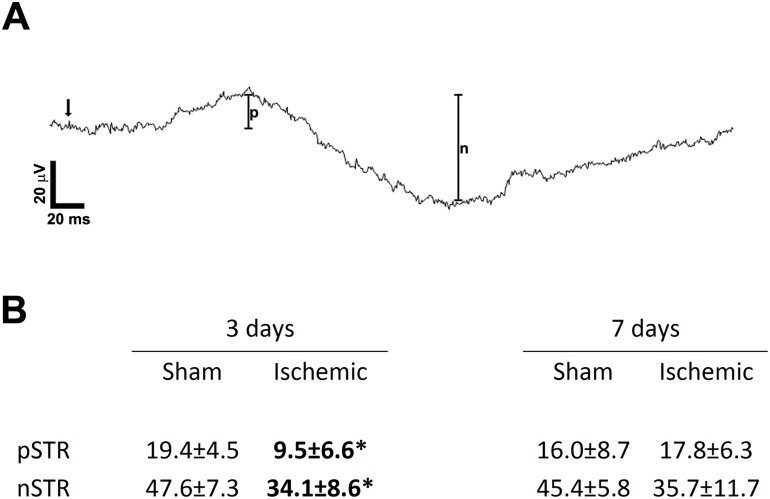
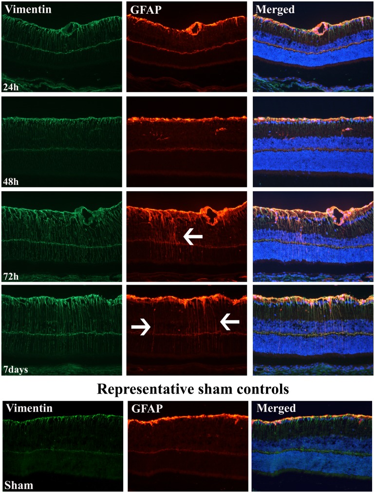
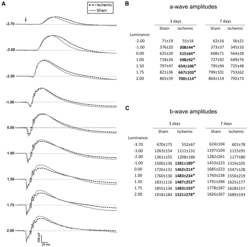
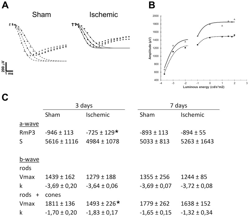
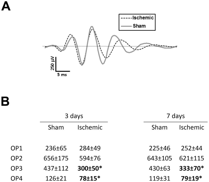
Cerebral vasculature is often the target of stroke studies. However, the vasculature supplying the eye might also be affected by ischemia. The aim of the present study was to investigate if the transient global cerebral ischemia (GCI) enhances vascular effect of endothelin-1 (ET-1) and 5-hydroxytryptamine/serotonin (5-HT) on the ophthalmic artery in rats, leading to delayed retinal damage. This was preformed using myography on the ophthalmic artery, coupled with immunohistochemistry and electroretinogram (ERG) to assess the ischemic consequences on the retina. Results showed a significant increase of ET-1 mediated vasoconstriction at 48 hours post ischemia. The retina did not exhibit any morphological changes throughout the study. However, we found an increase of GFAP and vimentin expression at 72 hours and 7 days after ischemia, indicating Müller cell mediated gliosis. ERG revealed significantly decreased function at 72 hours, but recovered almost completely after 7 days. In conclusion, we propose that the increased contractile response via ET-1 receptors in the ophthalmic artery after 48 hours may elicit negative retinal consequences due to a second ischemic period. This may exacerbate retinal damage after ischemia as illustrated by the decreased retinal function and Müller cell activation. The ophthalmic artery and ET-1 mediated vasoconstriction may be a valid and novel therapeutic target after longer periods of ischemic insults.
脑血管系统常常是中风研究的目标。然而,供应眼睛的血管系统也可能受到缺血的影响。本研究的目的是调查短暂性全脑缺血(GCI)是否会增强内皮素-1(ET-1)和5-羟色胺/血清素(5-HT)对大鼠眼动脉的血管效应,从而导致延迟性视网膜损伤。这是通过对眼动脉进行肌动描记法,并结合免疫组织化学和视网膜电图(ERG)来评估对视网膜的缺血后果来完成的。结果显示,缺血后48小时ET-1介导的血管收缩显著增加。在整个研究过程中,视网膜未表现出任何形态学变化。然而,我们发现在缺血后72小时和7天时,胶质纤维酸性蛋白(GFAP)和波形蛋白的表达增加,表明存在穆勒细胞介导的神经胶质增生。ERG显示在72小时时功能显著下降,但在7天后几乎完全恢复。总之,我们提出,48小时后眼动脉中通过ET-1受体增加的收缩反应可能由于第二个缺血期而引发负面的视网膜后果。这可能会加剧缺血后的视网膜损伤,如视网膜功能下降和穆勒细胞激活所示。眼动脉和ET-1介导的血管收缩可能是长时间缺血性损伤后的一个有效且新颖的治疗靶点。