Diana Angela, Wang Lai Mun, D'Costa Zenobia, Allen Paul, Azad Abul, Silva Michael A, Soonawalla Zahir, Liu Stanley, McKenna W Gillies, Muschel Ruth J, Fokas Emmanouil
Department of Oncology, CRUK/MRC Oxford Institute for Radiation Oncology, University of Oxford, Oxford, UK.
Department of Pathology, Oxford University Hospital NHS Foundation Trust, Oxford, UK.
Oncotarget. 2016 Jul 5;7(27):40992-41004. doi: 10.18632/oncotarget.10038.
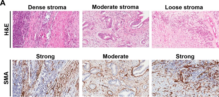
We examined the prognostic value of programmed cell death-1 (PD-1) and its ligand (PD-L1) together with CD8+ tumor-infiltrating lymphocytes (TILs) and FOXP3+ Tregs in resectable pancreatic ductal adenocarcinoma (PDAC) samples treated with adjuvant chemotherapy. Whole-mount FFPE tissue sections from 145 pancreatectomies were immunohistochemically stained for PD-1, PD-L1, CD8 and FOXP3. Their expression was correlated with clinicopathological characteristics, and overall survival (OS), progression-free survival (PFS), local progression-free survival (LPFS) and distant metastases free-survival (DMFS), in the context of stroma density (haematoxylin-eosin) and activity (alpha-smooth muscle actin) and in regard to intratumoral lymphoid aggregates. The median OS was 21 months after a mean follow-up of 20 months (range, 2-69 months). In multivariate analysis, high PD-1+ TILs expression was associated with better OS (p = 0.049), LPFS (p = 0.017) and DMFS (p = 0.021). Similar findings were observed for CD8+ TILs, whereas FOXP3 and PD-L1 lacked prognostic significance. Although TIL distribution was heterogeneous, tumors of high stroma density had higher infiltration of CD8+ TILs than loose density stroma and vice versa (p < 0.001), whereas no correlation was found with stromal activity. Sixty (41.4%) tumors contained lymphoid aggregates and the presence of PD-1+ TILs was associated with better OS (p = 0.030), LPFS (p = 0.025) and DMFS (p = 0.033), whereas CD8+ TILs only correlated with superior LPFS (p = 0.039). PD-1+ and CD8+ TILs constitute independent prognostic markers in patients with PDAC treated with adjuvant chemotherapy. Our study provides important insight on the role of PD-1/PD-L1 in the context of desmoplastic stroma and could help guide future immunotherapies in PDAC.
我们研究了程序性细胞死亡蛋白1(PD-1)及其配体(PD-L1)、CD8+肿瘤浸润淋巴细胞(TILs)和FOXP3+调节性T细胞(Tregs)在接受辅助化疗的可切除胰腺导管腺癌(PDAC)样本中的预后价值。对145例胰腺切除术的全层福尔马林固定石蜡包埋(FFPE)组织切片进行PD-1、PD-L1、CD8和FOXP3的免疫组织化学染色。在考虑基质密度(苏木精-伊红染色)和活性(α-平滑肌肌动蛋白)以及肿瘤内淋巴样聚集物的情况下,将它们的表达与临床病理特征、总生存期(OS)、无进展生存期(PFS)、局部无进展生存期(LPFS)和无远处转移生存期(DMFS)相关联。平均随访20个月(范围2 - 69个月)后,中位OS为21个月。在多变量分析中,高PD-1+TILs表达与更好的OS(p = 0.049)、LPFS(p = 0.017)和DMFS(p = 0.021)相关。CD8+TILs也观察到类似结果,而FOXP3和PD-L1缺乏预后意义。尽管TIL分布不均一,但高基质密度肿瘤比低基质密度肿瘤有更高的CD8+TILs浸润(p < 0.001),而与基质活性无相关性。60例(41.4%)肿瘤含有淋巴样聚集物,PD-1+TILs的存在与更好的OS(p = 0.030)、LPFS(p = 0.025)和DMFS(p = 0.033)相关,而CD8+TILs仅与较好的LPFS相关(p = 0.039)。在接受辅助化疗的PDAC患者中,PD-1+和CD8+TILs构成独立的预后标志物。我们的研究为PD-1/PD-L1在促纤维增生性基质背景下的作用提供了重要见解,并有助于指导未来PDAC的免疫治疗。