Department of Pancreatic Surgery, Fudan University Shanghai Cancer Center, Shanghai, China.
Department of Oncology Shanghai Medical College, Fudan University, Shanghai, China.
J Immunother Cancer. 2019 Aug 29;7(1):233. doi: 10.1186/s40425-019-0703-0.
Programmed cell death protein 1 (PD-1) is a key immune checkpoint that regulates peripheral tolerance and protects against autoimmunity. Programmed death ligand-2 (PD-L2) is a less studied ligand to PD-1 and has yet to be fully explored, especially in pancreatic ductal adenocarcinoma (PDAC).
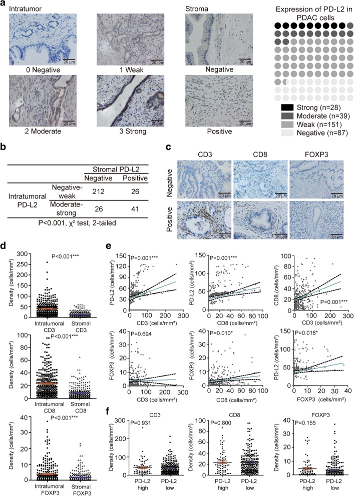
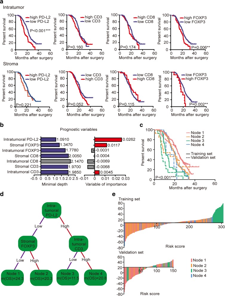
In this study, we performed immunohistochemistry to detect the PD-L2, CD3, CD8, transforming growth factor-β2 (TGF-β2) and FOXP3 levels in paraffin sections from 305 patients with resected PDAC as a training set. Expression levels of intratumoral and stromal immune markers were compared in relation to survival using Kaplan-Meier curves, random survival forest model and survival tree analysis. A multivariable Cox proportional-hazards model of associated markers was used to calculate the risk scores.
PD-L2 was expressed in 71.5% of PDAC samples and showed strong correlations with CD3+, CD8+ T cells and FOXP3+ regulatory T cell densities. High levels of intratumoral PD-L2 and FOXP3 were related to poor survival; only stromal FOXP3 overexpression was associated with worse prognosis. Four patterns generated from survival tree analysis demonstrated that PD-L2FOXP3 patients had the longest survival, while PD-L2CD3 patients had the shortest survival (P < 0.001). The area under the curve was 0.631(95% confidence interval (CI): 0.447-0.826) for the immune marker-based signature and 0.549 (95% CI: 0.323-0.829; P < 0.001) for the clinical parameter-based signature, which was consistent with the results in the validation set including 150 patients (P < 0.001). A higher risk score indicated shorter survival and could serve as an independent prognostic factor. PD-L2 was also showed associated with TGF-β2 and other immune molecules based on bioinformatics analysis.
Our work highlighted PD-L2 as a promising immunotherapeutic target with prognostic value combined with complex tumor infiltrating cells in PDAC.
程序性死亡蛋白 1(PD-1)是一种关键的免疫检查点,调节外周耐受并防止自身免疫。程序性死亡配体-2(PD-L2)是 PD-1 的一种研究较少的配体,尚未得到充分探索,特别是在胰腺导管腺癌(PDAC)中。
在这项研究中,我们使用免疫组织化学方法检测了 305 例接受手术切除的 PDAC 患者石蜡切片中的 PD-L2、CD3、CD8、转化生长因子-β2(TGF-β2)和 FOXP3 水平作为训练集。使用 Kaplan-Meier 曲线、随机生存森林模型和生存树分析比较了肿瘤内和基质免疫标志物与生存的关系。使用多变量 Cox 比例风险模型计算相关标志物的风险评分。
PD-L2 在 71.5%的 PDAC 样本中表达,并与 CD3+、CD8+T 细胞和 FOXP3+调节性 T 细胞密度呈强相关。肿瘤内高水平的 PD-L2 和 FOXP3 与生存不良相关;只有基质 FOXP3 过表达与预后不良相关。生存树分析生成的四种模式表明,PD-L2+FOXP3+患者的生存时间最长,而 PD-L2+CD3+患者的生存时间最短(P<0.001)。基于免疫标志物的特征曲线下面积为 0.631(95%置信区间(CI):0.447-0.826),基于临床参数的特征曲线下面积为 0.549(95%CI:0.323-0.829;P<0.001),在包括 150 例患者的验证集中得到了一致的结果(P<0.001)。较高的风险评分表明生存时间较短,可作为独立的预后因素。基于生物信息学分析,PD-L2 还与 TGF-β2 和其他免疫分子相关。
我们的工作强调了 PD-L2 作为一种有前途的免疫治疗靶点,具有与 PDAC 中复杂肿瘤浸润细胞结合的预后价值。