Pollard Amelia Kate, Craig Emma Louise, Chakrabarti Lisa
School of Veterinary Medicine and Science, University of Nottingham, Sutton Bonington, United Kingdom.
PLoS One. 2016 Jun 22;11(6):e0157405. doi: 10.1371/journal.pone.0157405. eCollection 2016.
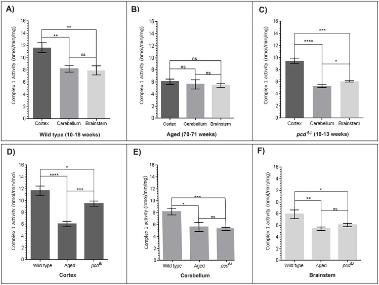
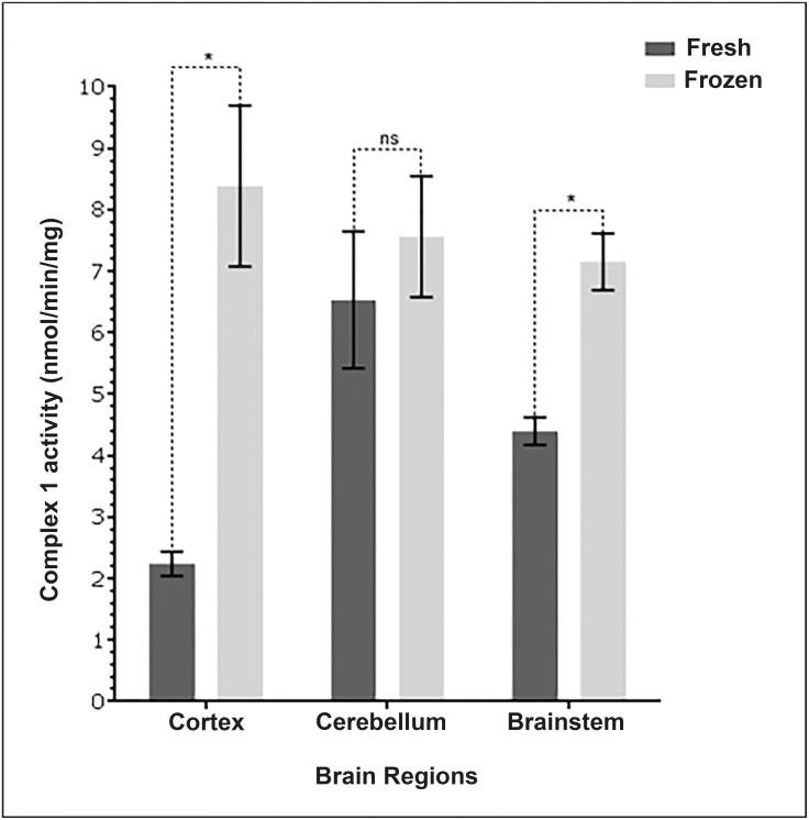
Mitochondrial function, in particular complex 1 of the electron transport chain (ETC), has been shown to decrease during normal ageing and in neurodegenerative disease. However, there is some debate concerning which area of the brain has the greatest complex 1 activity. It is important to identify the pattern of activity in order to be able to gauge the effect of age or disease related changes. We determined complex 1 activity spectrophotometrically in the cortex, brainstem and cerebellum of middle aged mice (70-71 weeks), a cerebellar ataxic neurodegeneration model (pcd5J) and young wild type controls. We share our updated protocol on the measurements of complex1 activity and find that mitochondrial fractions isolated from frozen tissues can be measured for robust activity. We show that complex 1 activity is clearly highest in the cortex when compared with brainstem and cerebellum (p<0.003). Cerebellum and brainstem mitochondria exhibit similar levels of complex 1 activity in wild type brains. In the aged brain we see similar levels of complex 1 activity in all three-brain regions. The specific activity of complex 1 measured in the aged cortex is significantly decreased when compared with controls (p<0.0001). Both the cerebellum and brainstem mitochondria also show significantly reduced activity with ageing (p<0.05). The mouse model of ataxia predictably has a lower complex 1 activity in the cerebellum, and although reductions are measured in the cortex and brain stem, the remaining activity is higher than in the aged brains. We present clear evidence that complex 1 activity decreases across the brain with age and much more specifically in the cerebellum of the pcd5j mouse. Mitochondrial impairment can be a region specific phenomenon in disease, but in ageing appears to affect the entire brain, abolishing the pattern of higher activity in cortical regions.
线粒体功能,尤其是电子传递链(ETC)的复合体I,已被证明在正常衰老过程和神经退行性疾病中会下降。然而,关于大脑的哪个区域具有最高的复合体I活性存在一些争议。确定活性模式对于能够评估年龄或疾病相关变化的影响很重要。我们用分光光度法测定了中年小鼠(70 - 71周)、小脑性共济失调神经退行性模型(pcd5J)和年轻野生型对照小鼠的皮质、脑干和小脑中复合体I的活性。我们分享了关于复合体I活性测量的更新方案,并发现从冷冻组织中分离的线粒体组分可以测量出强劲的活性。我们表明,与脑干和小脑相比,复合体I活性在皮质中明显最高(p<0.003)。在野生型大脑中,小脑和脑干线粒体表现出相似水平的复合体I活性。在老年大脑中,我们在所有三个脑区都看到了相似水平的复合体I活性。与对照组相比,老年皮质中测得的复合体I比活性显著降低(p<0.0001)。小脑和脑干线粒体的活性也随着衰老而显著降低(p<0.05)。共济失调小鼠模型可预测地在小脑中具有较低的复合体I活性,并且尽管在皮质和脑干中测量到了活性降低,但剩余活性高于老年大脑。我们提供了明确的证据,表明复合体I活性随着年龄增长在整个大脑中下降,并且在pcd5j小鼠的小脑中下降得更明显。线粒体损伤在疾病中可能是一种区域特异性现象,但在衰老过程中似乎会影响整个大脑,消除皮质区域较高活性的模式。