Yuan Jing, Yu Jian-Xiong
Eye Center, Renmin Hospital of Wuhan University, Wuhan, Hubei Province, China.
Department of Gastrointestinal Surgery, Renmin Hospital of Wuhan University, Wuhan, Hubei Province, China.
Neural Regen Res. 2016 May;11(5):846-53. doi: 10.4103/1673-5374.182764.
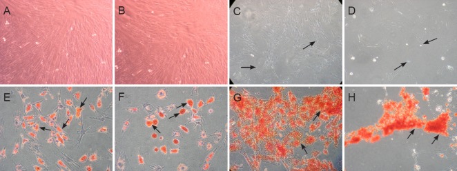
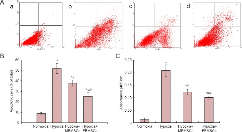
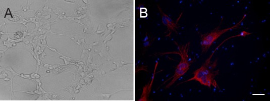
Bone marrow mesenchymal stem cells can reduce retinal ganglion cell death and effectively prevent vision loss. Previously, we found that during differentiation, female rhesus monkey bone marrow mesenchymal stem cells acquire a higher neurogenic potential compared with male rhesus monkey bone marrow mesenchymal stem cells. This suggests that female bone marrow mesenchymal stem cells have a stronger neuroprotective effect than male bone marrow mesenchymal stem cells. Here, we first isolated and cultured bone marrow mesenchymal stem cells from female and male rats by density gradient centrifugation. Retinal tissue from newborn rats was prepared by enzymatic digestion to obtain primary retinal ganglion cells. Using the transwell system, retinal ganglion cells were co-cultured with bone marrow mesenchymal stem cells under hypoxia. Cell apoptosis was detected by flow cytometry and caspase-3 activity assay. We found a marked increase in apoptotic rate and caspase-3 activity of retinal ganglion cells after 24 hours of hypoxia compared with normoxia. Moreover, apoptotic rate and caspase-3 activity of retinal ganglion cells significantly decreased with both female and male bone marrow mesenchymal stem cell co-culture under hypoxia compared with culture alone, with more significant effects from female bone marrow mesenchymal stem cells. Our results indicate that bone marrow mesenchymal stem cells exert a neuroprotective effect against hypoxia-induced apoptosis of retinal ganglion cells, and also that female cells have greater neuroprotective ability compared with male cells.
骨髓间充质干细胞可减少视网膜神经节细胞死亡,并有效预防视力丧失。此前,我们发现,在分化过程中,雌性恒河猴骨髓间充质干细胞比雄性恒河猴骨髓间充质干细胞具有更高的神经发生潜能。这表明雌性骨髓间充质干细胞比雄性骨髓间充质干细胞具有更强的神经保护作用。在此,我们首先通过密度梯度离心法从雌性和雄性大鼠中分离并培养骨髓间充质干细胞。通过酶消化制备新生大鼠的视网膜组织以获得原代视网膜神经节细胞。使用transwell系统,在缺氧条件下将视网膜神经节细胞与骨髓间充质干细胞共培养。通过流式细胞术和caspase-3活性测定检测细胞凋亡。我们发现,与常氧相比,缺氧24小时后视网膜神经节细胞的凋亡率和caspase-3活性显著增加。此外,与单独培养相比,在缺氧条件下雌性和雄性骨髓间充质干细胞共培养时,视网膜神经节细胞的凋亡率和caspase-3活性均显著降低,雌性骨髓间充质干细胞的作用更显著。我们的结果表明,骨髓间充质干细胞对缺氧诱导的视网膜神经节细胞凋亡具有神经保护作用,并且雌性细胞比雄性细胞具有更强的神经保护能力。