• 文献检索
  • 文档翻译
  • 深度研究
  • 学术资讯
  • Suppr Zotero 插件Zotero 插件
  • 邀请有礼
  • 套餐&价格
  • 历史记录
应用&插件
Suppr Zotero 插件Zotero 插件浏览器插件Mac 客户端Windows 客户端微信小程序
定价
高级版会员购买积分包购买API积分包
服务
文献检索文档翻译深度研究API 文档MCP 服务
关于我们
关于 Suppr公司介绍联系我们用户协议隐私条款
关注我们

Suppr 超能文献

核心技术专利:CN118964589B侵权必究
粤ICP备2023148730 号-1Suppr @ 2026

文献检索

告别复杂PubMed语法,用中文像聊天一样搜索,搜遍4000万医学文献。AI智能推荐,让科研检索更轻松。

立即免费搜索

文件翻译

保留排版,准确专业,支持PDF/Word/PPT等文件格式,支持 12+语言互译。

免费翻译文档

深度研究

AI帮你快速写综述,25分钟生成高质量综述,智能提取关键信息,辅助科研写作。

立即免费体验

视神经挤压伤及细胞治疗后间充质干细胞的分布及其对神经元存活和轴突再生的影响

Distribution of mesenchymal stem cells and effects on neuronal survival and axon regeneration after optic nerve crush and cell therapy.

作者信息

Mesentier-Louro Louise Alessandra, Zaverucha-do-Valle Camila, da Silva-Junior Almir Jordão, Nascimento-Dos-Santos Gabriel, Gubert Fernanda, de Figueirêdo Ana Beatriz Padilha, Torres Ana Luiza, Paredes Bruno D, Teixeira Camila, Tovar-Moll Fernanda, Mendez-Otero Rosalia, Santiago Marcelo F

机构信息

Instituto de Biofísica Carlos Chagas Filho, Universidade Federal do Rio de Janeiro, Rio de Janeiro, Brazil; Instituto Nacional de Ciência e Tecnologia de Biologia Estrutural e Bioimagem, INBEB, Rio de Janeiro, Brazil.

National Center of Structural Biology and Bioimaging (CENABIO), Universidade Federal do Rio de Janeiro, Rio de Janeiro, Brazil.

出版信息

PLoS One. 2014 Oct 27;9(10):e110722. doi: 10.1371/journal.pone.0110722. eCollection 2014.

DOI:10.1371/journal.pone.0110722
PMID:25347773
原文链接:https://pmc.ncbi.nlm.nih.gov/articles/PMC4210195/
Abstract

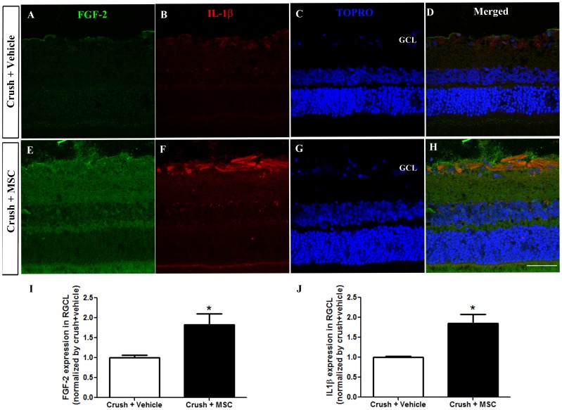
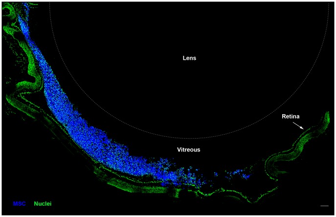
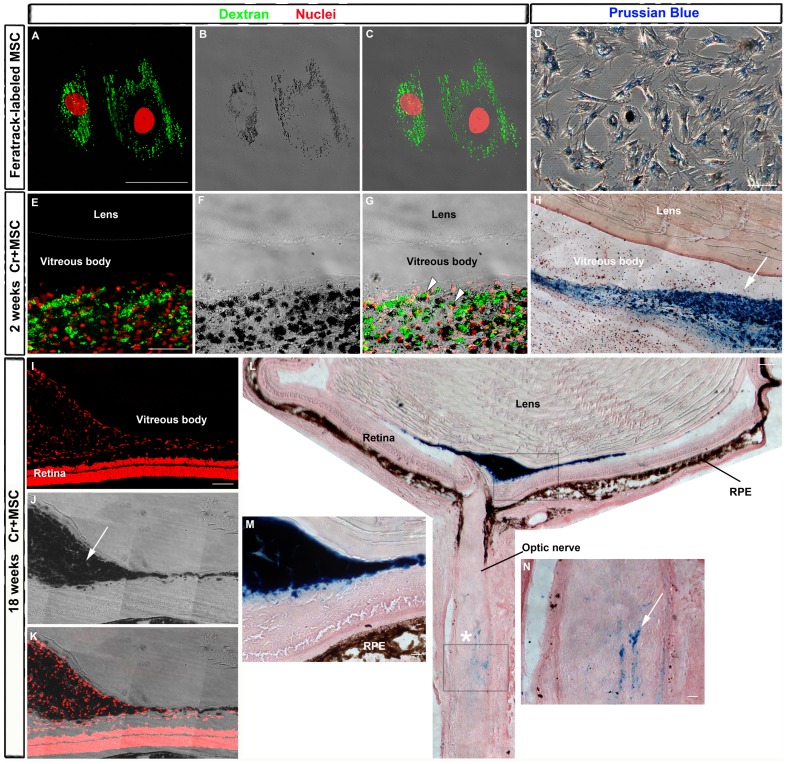
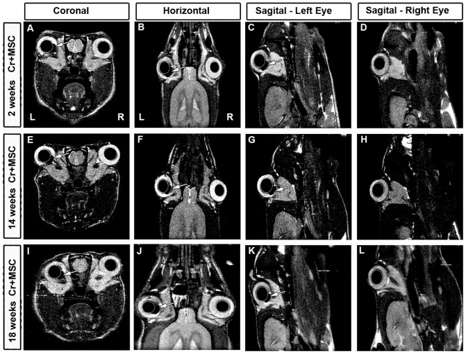
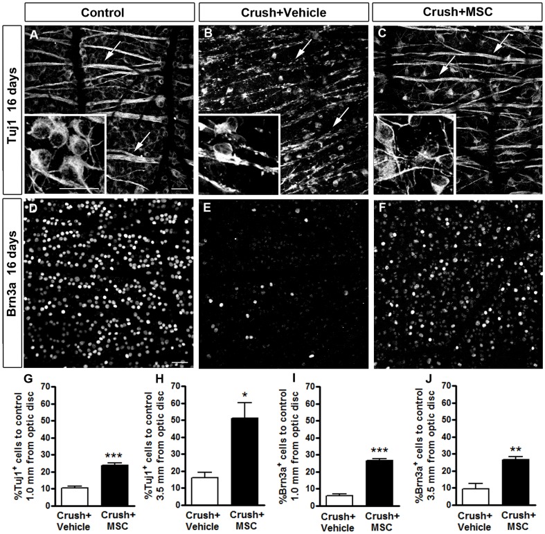
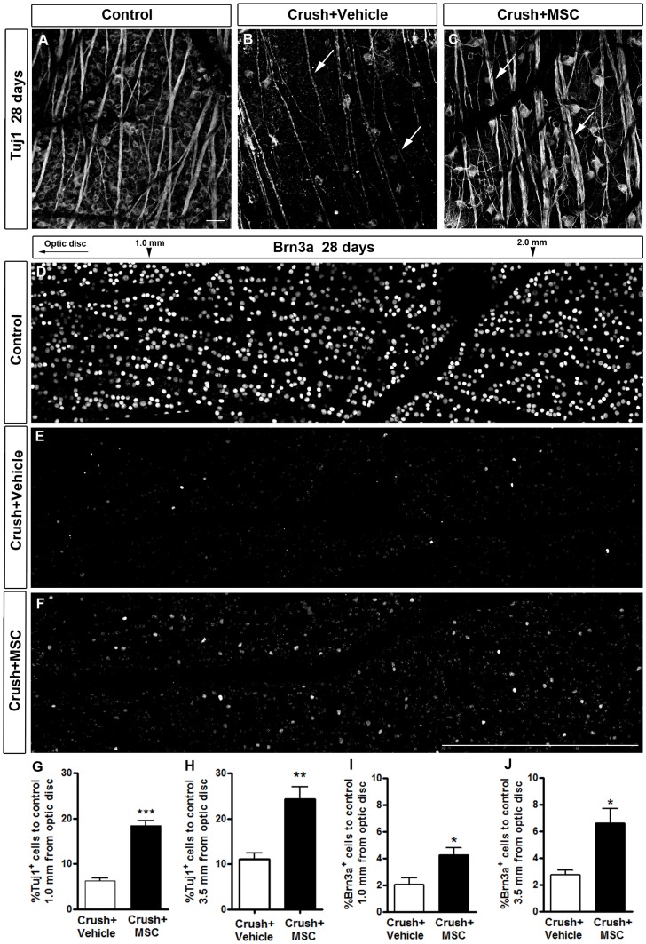
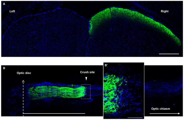
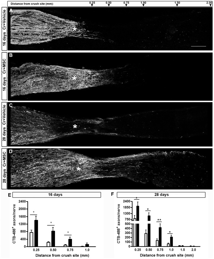
Bone marrow-derived cells have been used in different animal models of neurological diseases. We investigated the therapeutic potential of mesenchymal stem cells (MSC) injected into the vitreous body in a model of optic nerve injury. Adult (3-5 months old) Lister Hooded rats underwent unilateral optic nerve crush followed by injection of MSC or the vehicle into the vitreous body. Before they were injected, MSC were labeled with a fluorescent dye or with superparamagnetic iron oxide nanoparticles, which allowed us to track the cells in vivo by magnetic resonance imaging. Sixteen and 28 days after injury, the survival of retinal ganglion cells was evaluated by assessing the number of Tuj1- or Brn3a-positive cells in flat-mounted retinas, and optic nerve regeneration was investigated after anterograde labeling of the optic axons with cholera toxin B conjugated to Alexa 488. Transplanted MSC remained in the vitreous body and were found in the eye for several weeks. Cell therapy significantly increased the number of Tuj1- and Brn3a-positive cells in the retina and the number of axons distal to the crush site at 16 and 28 days after optic nerve crush, although the RGC number decreased over time. MSC therapy was associated with an increase in the FGF-2 expression in the retinal ganglion cells layer, suggesting a beneficial outcome mediated by trophic factors. Interleukin-1β expression was also increased by MSC transplantation. In summary, MSC protected RGC and stimulated axon regeneration after optic nerve crush. The long period when the transplanted cells remained in the eye may account for the effect observed. However, further studies are needed to overcome eventually undesirable consequences of MSC transplantation and to potentiate the beneficial ones in order to sustain the neuroprotective effect overtime.

摘要

骨髓来源的细胞已被用于多种神经疾病的动物模型。我们在视神经损伤模型中研究了注入玻璃体的间充质干细胞(MSC)的治疗潜力。成年(3 - 5个月大)的利斯特戴帽大鼠接受单侧视神经挤压,随后将MSC或赋形剂注入玻璃体。在注射前,MSC用荧光染料或超顺磁性氧化铁纳米颗粒标记,这使我们能够通过磁共振成像在体内追踪细胞。损伤后16天和28天,通过评估平铺视网膜中Tuj1或Brn3a阳性细胞的数量来评估视网膜神经节细胞的存活情况,并在用与Alexa 488偶联的霍乱毒素B对视神经轴突进行顺行标记后研究视神经再生。移植的MSC留在玻璃体内,并在眼中存在数周。细胞治疗在视神经挤压后16天和28天显著增加了视网膜中Tuj1和Brn3a阳性细胞的数量以及挤压部位远端的轴突数量,尽管视网膜神经节细胞数量随时间减少。MSC治疗与视网膜神经节细胞层中FGF - 2表达增加有关,提示由营养因子介导的有益结果。MSC移植也增加了白细胞介素 - 1β的表达。总之,MSC在视神经挤压后保护了视网膜神经节细胞并刺激了轴突再生。移植细胞在眼中停留的较长时间可能解释了观察到的效果。然而,需要进一步研究以最终克服MSC移植的不良后果并增强有益效果,以便长期维持神经保护作用。

https://cdn.ncbi.nlm.nih.gov/pmc/blobs/2852/4210195/b1f8861bd1a8/pone.0110722.g010.jpg
https://cdn.ncbi.nlm.nih.gov/pmc/blobs/2852/4210195/a5bd6912b787/pone.0110722.g001.jpg
https://cdn.ncbi.nlm.nih.gov/pmc/blobs/2852/4210195/3875416e1ddd/pone.0110722.g002.jpg
https://cdn.ncbi.nlm.nih.gov/pmc/blobs/2852/4210195/29f0753d02df/pone.0110722.g003.jpg
https://cdn.ncbi.nlm.nih.gov/pmc/blobs/2852/4210195/8103ac1521c0/pone.0110722.g004.jpg
https://cdn.ncbi.nlm.nih.gov/pmc/blobs/2852/4210195/c1841ac211e2/pone.0110722.g005.jpg
https://cdn.ncbi.nlm.nih.gov/pmc/blobs/2852/4210195/c7465b6379fc/pone.0110722.g006.jpg
https://cdn.ncbi.nlm.nih.gov/pmc/blobs/2852/4210195/00150a3259c3/pone.0110722.g007.jpg
https://cdn.ncbi.nlm.nih.gov/pmc/blobs/2852/4210195/6eb9a63bb8cd/pone.0110722.g008.jpg
https://cdn.ncbi.nlm.nih.gov/pmc/blobs/2852/4210195/225debccf1c9/pone.0110722.g009.jpg
https://cdn.ncbi.nlm.nih.gov/pmc/blobs/2852/4210195/b1f8861bd1a8/pone.0110722.g010.jpg

相似文献

1
Distribution of mesenchymal stem cells and effects on neuronal survival and axon regeneration after optic nerve crush and cell therapy.视神经挤压伤及细胞治疗后间充质干细胞的分布及其对神经元存活和轴突再生的影响
PLoS One. 2014 Oct 27;9(10):e110722. doi: 10.1371/journal.pone.0110722. eCollection 2014.
2
Intravitreally transplanted dental pulp stem cells promote neuroprotection and axon regeneration of retinal ganglion cells after optic nerve injury.玻璃体内移植牙髓干细胞促进视神经损伤后视网膜神经节细胞的神经保护和轴突再生。
Invest Ophthalmol Vis Sci. 2013 Nov 15;54(12):7544-56. doi: 10.1167/iovs.13-13045.
3
Long-term neuronal survival, regeneration, and transient target reconnection after optic nerve crush and mesenchymal stem cell transplantation.视神经挤压和间充质干细胞移植后神经元的长期存活、再生和短暂的靶重新连接。
Stem Cell Res Ther. 2019 Apr 17;10(1):121. doi: 10.1186/s13287-019-1226-9.
4
Sustained effect of bone marrow mononuclear cell therapy in axonal regeneration in a model of optic nerve crush.骨髓单个核细胞疗法对视神经挤压模型中轴突再生的持续作用。
Brain Res. 2014 Oct 31;1587:54-68. doi: 10.1016/j.brainres.2014.08.070. Epub 2014 Sep 6.
5
AAV-mediated expression of CNTF promotes long-term survival and regeneration of adult rat retinal ganglion cells.腺相关病毒介导的睫状神经营养因子表达促进成年大鼠视网膜神经节细胞的长期存活和再生。
Gene Ther. 2006 Sep;13(18):1328-41. doi: 10.1038/sj.gt.3302791. Epub 2006 May 18.
6
Human umbilical cord blood mononuclear cells and chorionic plate-derived mesenchymal stem cells promote axon survival in a rat model of optic nerve crush injury.人脐带血单个核细胞和绒毛膜板来源的间充质干细胞在大鼠视神经挤压伤模型中促进轴突存活。
Int J Mol Med. 2016 May;37(5):1170-80. doi: 10.3892/ijmm.2016.2532. Epub 2016 Mar 17.
7
Immunohistochemical localization of CNTFRalpha in adult mouse retina and optic nerve following intraorbital nerve crush: evidence for the axonal loss of a trophic factor receptor after injury.眶内神经挤压后成年小鼠视网膜和视神经中CNTFRalpha的免疫组织化学定位:损伤后营养因子受体轴突损失的证据。
J Comp Neurol. 2007 Jan 10;500(2):384-400. doi: 10.1002/cne.21174.
8
Effects of a combinatorial treatment with gene and cell therapy on retinal ganglion cell survival and axonal outgrowth after optic nerve injury.基因和细胞联合治疗对视神经损伤后视网膜神经节细胞存活和轴突生长的影响。
Gene Ther. 2020 Feb;27(1-2):27-39. doi: 10.1038/s41434-019-0089-0. Epub 2019 Jun 26.
9
Topical administration of a Rock/Net inhibitor promotes retinal ganglion cell survival and axon regeneration after optic nerve injury.局部应用Rock/Net抑制剂可促进视神经损伤后视网膜神经节细胞存活和轴突再生。
Exp Eye Res. 2017 May;158:33-42. doi: 10.1016/j.exer.2016.07.006. Epub 2016 Jul 18.
10
Effects of a conventional photocoagulator and a 3-ns pulse laser on preconditioning responses and retinal ganglion cell survival after optic nerve crush.传统光凝器和3纳秒脉冲激光对视神经挤压后预处理反应及视网膜神经节细胞存活的影响。
Exp Eye Res. 2014 Oct;127:77-90. doi: 10.1016/j.exer.2014.07.008. Epub 2014 Jul 21.

引用本文的文献

1
Extracellular Vesicle Metabolomics Holds Promise for Adult Axon Regeneration.细胞外囊泡代谢组学有望促进成体轴突再生。
Metabolites. 2025 Jul 4;15(7):454. doi: 10.3390/metabo15070454.
2
Potential therapeutic applications of stem cells in animal models of ocular affections.干细胞在眼部疾病动物模型中的潜在治疗应用。
Inflamm Regen. 2025 Jul 21;45(1):23. doi: 10.1186/s41232-025-00380-7.
3
Rekindling Vision: Innovative Strategies for Treating Retinal Degeneration.重燃光明:治疗视网膜变性的创新策略

本文引用的文献

1
Neuronal STAT3 activation is essential for CNTF- and inflammatory stimulation-induced CNS axon regeneration.神经元 STAT3 的激活对于 CNTF 和炎症刺激诱导的中枢神经系统轴突再生是必不可少的。
Cell Death Dis. 2013 Sep 19;4(9):e805. doi: 10.1038/cddis.2013.310.
2
Neutrophils express oncomodulin and promote optic nerve regeneration.中性粒细胞表达骨桥蛋白并促进视神经再生。
J Neurosci. 2013 Sep 11;33(37):14816-24. doi: 10.1523/JNEUROSCI.5511-12.2013.
3
Interleukin-1β orchestrates underlying inflammatory responses in microglia via Krüppel-like factor 4.
Int J Mol Sci. 2025 Apr 25;26(9):4078. doi: 10.3390/ijms26094078.
4
Restorative potential of ciliary body cells in a retinal ganglion cell degeneration model.视网膜神经节细胞变性模型中睫状体细胞的修复潜力
Sci Rep. 2025 May 3;15(1):15503. doi: 10.1038/s41598-025-00283-0.
5
Growth Hormone Neuroprotective Effects After an Optic Nerve Crush in the Male Rat.雄性大鼠视神经挤压后生长激素的神经保护作用。
Invest Ophthalmol Vis Sci. 2024 Nov 4;65(13):17. doi: 10.1167/iovs.65.13.17.
6
Concept of Normativity in Multi-Omics Analysis of Axon Regeneration.轴突再生的多组学分析中的规范性概念。
Biomolecules. 2024 Jun 21;14(7):735. doi: 10.3390/biom14070735.
7
Mesenchymal stem cells for repairing glaucomatous optic nerve.用于修复青光眼性视神经的间充质干细胞
Int J Ophthalmol. 2024 Apr 18;17(4):748-760. doi: 10.18240/ijo.2024.04.20. eCollection 2024.
8
Cell-free fat extract promotes axon regeneration and retinal ganglion cells survival in traumatic optic neuropathy.无细胞脂肪提取物促进外伤性视神经病变中的轴突再生和视网膜神经节细胞存活。
Front Cell Neurosci. 2024 Mar 7;18:1344853. doi: 10.3389/fncel.2024.1344853. eCollection 2024.
9
New Perspectives in Stem Cell Transplantation and Associated Therapies to Treat Retinal Diseases: From Gene Editing to 3D Bioprinting.干细胞移植与相关疗法在视网膜疾病治疗中的新视角:从基因编辑到 3D 生物打印。
Stem Cell Rev Rep. 2024 Apr;20(3):722-737. doi: 10.1007/s12015-024-10689-4. Epub 2024 Feb 6.
10
Interplay between mesenchymal stromal cells and the immune system after transplantation: implications for advanced cell therapy in the retina.移植后间充质基质细胞与免疫系统之间的相互作用:对视网膜高级细胞治疗的影响。
Neural Regen Res. 2024 Mar;19(3):542-547. doi: 10.4103/1673-5374.380876.
白细胞介素-1β 通过 Kruppel 样因子 4 来调控小胶质细胞中的潜在炎症反应。
J Neurochem. 2013 Oct;127(2):233-44. doi: 10.1111/jnc.12382. Epub 2013 Aug 19.
4
Interleukin-6 contributes to CNS axon regeneration upon inflammatory stimulation.白细胞介素-6 有助于炎症刺激后的中枢神经系统轴突再生。
Cell Death Dis. 2013 Apr 25;4(4):e609. doi: 10.1038/cddis.2013.126.
5
Towards clinical application of mesenchymal stem cells for treatment of neurological diseases of the central nervous system.间充质干细胞在中枢神经系统神经疾病治疗中的临床应用探索。
J Neuroimmune Pharmacol. 2013 Dec;8(5):1062-76. doi: 10.1007/s11481-013-9456-6. Epub 2013 Apr 12.
6
The rise of cell therapy trials for stroke: review of published and registered studies.细胞治疗卒中临床试验的兴起:已发表和注册研究的综述。
Stem Cells Dev. 2013 Aug 1;22(15):2095-111. doi: 10.1089/scd.2013.0089. Epub 2013 Apr 25.
7
Concise review: Current concepts in bone marrow microenvironmental regulation of hematopoietic stem and progenitor cells.简明综述:骨髓造血微环境调控造血干/祖细胞的最新概念。
Stem Cells. 2013 Jun;31(6):1044-50. doi: 10.1002/stem.1370.
8
The meaning, the sense and the significance: translating the science of mesenchymal stem cells into medicine.意义、概念和重要性:将间充质干细胞科学转化为医学。
Nat Med. 2013 Jan;19(1):35-42. doi: 10.1038/nm.3028. Epub 2013 Jan 7.
9
Combinatorial therapy stimulates long-distance regeneration, target reinnervation, and partial recovery of vision after optic nerve injury in mice.组合疗法可刺激视神经损伤后的小鼠进行长距离再生、靶神经再支配和部分视力恢复。
Int Rev Neurobiol. 2012;106:153-72. doi: 10.1016/B978-0-12-407178-0.00007-7.
10
Whole number, distribution and co-expression of brn3 transcription factors in retinal ganglion cells of adult albino and pigmented rats.成年白化和色素大鼠视网膜神经节细胞中 brn3 转录因子的总数、分布和共表达。
PLoS One. 2012;7(11):e49830. doi: 10.1371/journal.pone.0049830. Epub 2012 Nov 16.