Department of Ophthalmology, Xijing Hospital, Fourth Military Medical University, Xi'an 710032, China.
Department of Medical Genetics and Developmental Biology, Fourth Military Medical University, Xi'an 710032, China.
Sci Rep. 2016 Jun 24;6:28617. doi: 10.1038/srep28617.
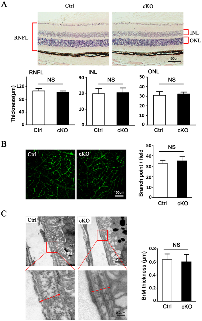
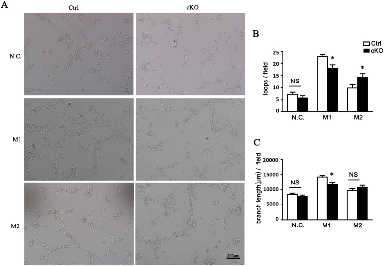
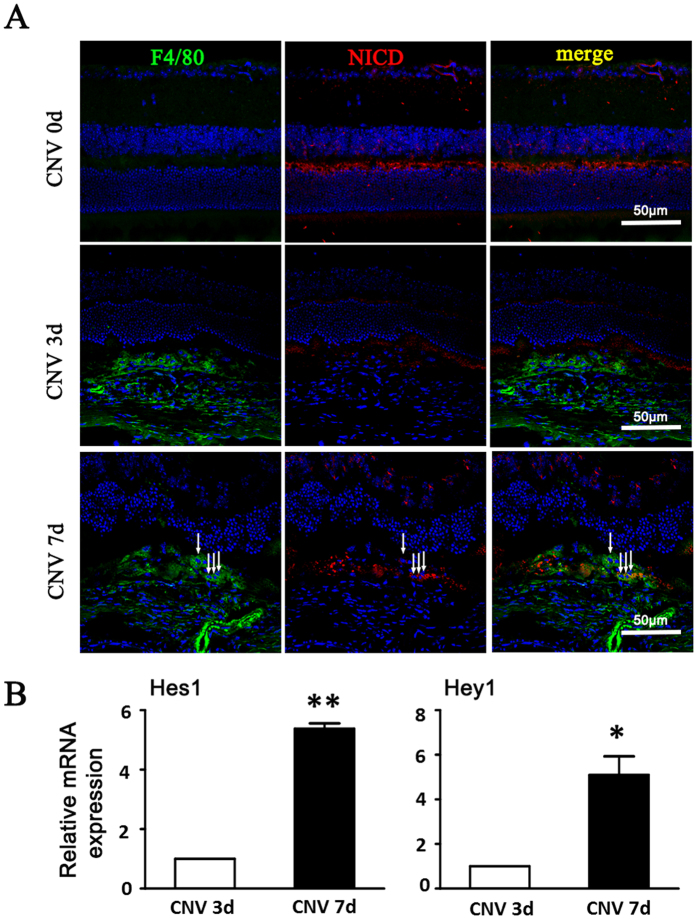
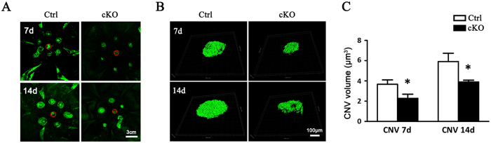
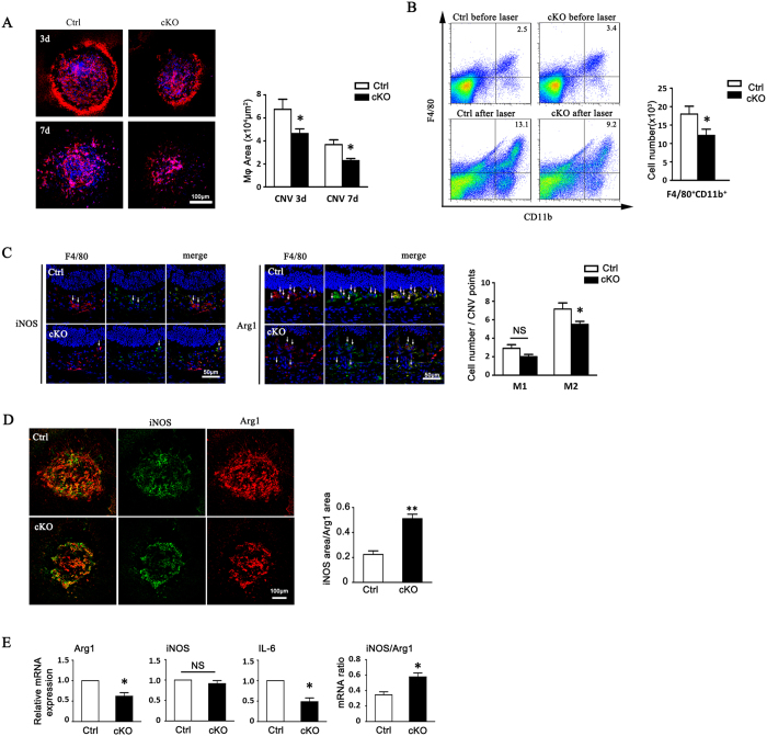
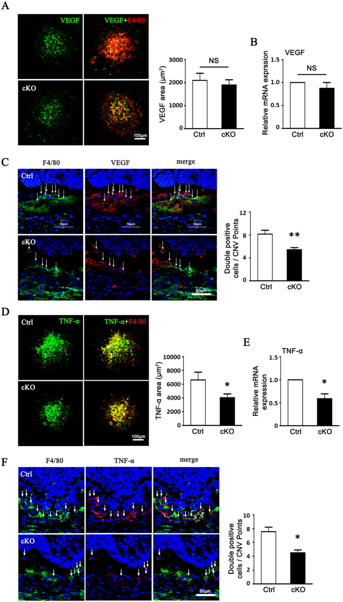
Macrophages have been recognized as an important inflammatory component in choroidal neovascularization (CNV). However, it is unclear how these cells are activated and polarized, how they affect angiogenesis and what the underlining mechanisms are during CNV. Notch signaling has been implicated in macrophage activation. Previously we have shown that inducible disruption of RBP-J, the critical transcription factor of Notch signaling, in adult mice results in enhanced CNV, but it is unclear what is the role of macrophage-specific Notch signaling in the development of CNV. In the current study, by using the myeloid specific RBP-J knockout mouse model combined with the laser-induced CNV model, we show that disruption of Notch signaling in macrophages displayed attenuated CNV growth, reduced macrophage infiltration and activation, and alleviated angiogenic response after laser induction. The inhibition of CNV occurred with reduced expression of VEGF and TNF-α in infiltrating inflammatory macrophages in myeloid specific RBP-J knockout mice. These changes might result in direct inhibition of EC lumen formation, as shown in an in vitro study. Therefore, clinical intervention of Notch signaling in CNV needs to pinpoint myeloid lineage to avoid the counteractive effects of global inhibition.
巨噬细胞已被认为是脉络膜新生血管(CNV)中的一个重要炎症成分。然而,目前尚不清楚这些细胞是如何被激活和极化的,它们如何影响血管生成,以及在 CNV 过程中潜在的机制是什么。Notch 信号通路已被证实与巨噬细胞的激活有关。之前我们的研究表明,在成年小鼠中诱导性破坏 Notch 信号通路的关键转录因子 RBP-J,会导致 CNV 增强,但巨噬细胞特异性 Notch 信号通路在 CNV 发展中的作用尚不清楚。在本研究中,我们使用髓系特异性 RBP-J 敲除小鼠模型结合激光诱导的 CNV 模型,发现巨噬细胞中 Notch 信号通路的破坏会导致 CNV 生长减弱、巨噬细胞浸润和激活减少,以及激光诱导后的血管生成反应减轻。髓系特异性 RBP-J 敲除小鼠中浸润性炎症巨噬细胞中 VEGF 和 TNF-α的表达减少,抑制了 CNV 的发生。这些变化可能导致 EC 管腔形成的直接抑制,如体外研究所示。因此,CNV 中 Notch 信号通路的临床干预需要针对髓系谱系,以避免全局性抑制的拮抗作用。