Department of Nuclear Medicine & PET Centre, Aarhus University Hospital, DK8000, Aarhus, Denmark.
Department of Radiology, Aarhus University Hospital, DK8000, Aarhus, Denmark.
EJNMMI Res. 2016 Dec;6(1):56. doi: 10.1186/s13550-016-0206-7. Epub 2016 Jun 24.
Positron emission tomography (PET) with the liver-specific galactose tracer 2-[(18)F]fluoro-2-deoxy-D-galactose ((18)F-FDGal) may improve diagnosis of hepatocellular carcinoma (HCC). The aim of this study was to test which of three different (18)F-FDGal PET protocols gives the highest tumour-to-background (T/B) ratio on PET images and thus better detection of HCC tumours.
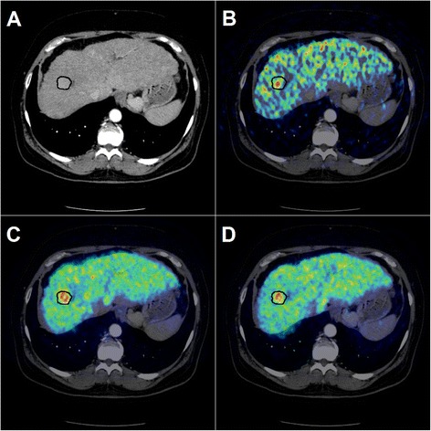
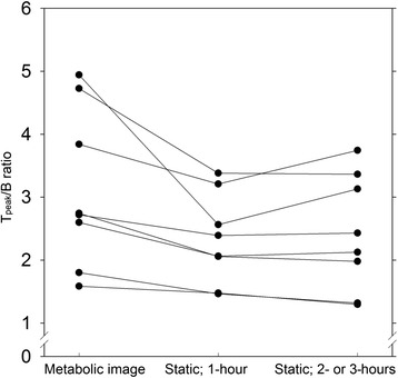
Ten patients with a total of 15 hepatic HCC tumours were enrolled prior to treatment. An experienced radiologist defined volumes of interest (VOIs) encircling HCC tumours on contrast-enhanced CT (ce-CT) images. Three PET/CT protocols were conducted following an intravenous (18)F-FDGal injection: (i) a 20-min dynamic PET/CT of the liver (to generate a 3D metabolic image), (ii) a traditional static whole-body PET/CT after 1 h, and (iii) a late static whole-body PET/CT after 2 or 3 h. PET images from each PET/CT protocol were fused with ce-CT images, and the average standardized uptake values (SUV) in tumour and background liver tissue were used to calculate (T/B) ratios. Furthermore, Tpeak/B ratios were calculated using the five hottest voxels in all hot tumours. The ratios for the three different PET protocols were compared.
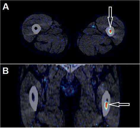
For the individual tumours, there was no significant difference in the T/B ratio between the three PET protocols. The metabolic image yielded higher Tpeak/B ratios than the two static images, but it was easier to identify tumours on the static images. One extrahepatic metastasis was detected.
Neither metabolic images nor static whole-body images acquired 2 or 3 h after (18)F-FDGal injection offered an advantage to traditional whole-body PET/CT images acquired after 1 h for detection of HCC.
正电子发射断层扫描(PET)使用肝脏特异性半乳糖示踪剂 2-[(18)F]氟-2-脱氧-D-半乳糖((18)F-FDGal)可提高肝细胞癌(HCC)的诊断水平。本研究旨在测试三种不同的(18)F-FDGal PET 方案中哪一种可在 PET 图像上获得最高的肿瘤与背景(T/B)比值,从而更好地检测 HCC 肿瘤。
在治疗前,共纳入了 10 名患有 15 个肝脏 HCC 肿瘤的患者。一位经验丰富的放射科医生在对比增强 CT(ce-CT)图像上定义了 HCC 肿瘤的感兴趣区(VOI)。在静脉注射(18)F-FDGal 后进行了三种 PET/CT 方案:(i)肝脏 20 分钟的动态 PET/CT(生成 3D 代谢图像),(ii)1 小时后的传统静态全身 PET/CT,以及(iii)2 或 3 小时后的延迟静态全身 PET/CT。从每个 PET/CT 方案获得的 PET 图像与 ce-CT 图像融合,使用肿瘤和背景肝组织的平均标准化摄取值(SUV)来计算(T/B)比值。此外,还使用所有热点肿瘤中的五个最热体素来计算 Tpeak/B 比值。比较了三种不同 PET 方案的比值。
对于单个肿瘤,三种 PET 方案之间的 T/B 比值没有显著差异。代谢图像产生的 Tpeak/B 比值高于两种静态图像,但在静态图像上更容易识别肿瘤。还检测到一个肝外转移灶。
对于 HCC 的检测,与 1 小时后采集的传统全身 PET/CT 图像相比,(18)F-FDGal 注射后 2 或 3 小时采集的代谢图像或全身静态图像均没有优势。