Department of Nuclear Medicine, Military Institute of Medicine, Warsaw, Poland.
Affidea Mazovian PET/CT Medical Centre, Warsaw, Poland.
PLoS One. 2021 Feb 10;16(2):e0246848. doi: 10.1371/journal.pone.0246848. eCollection 2021.
We aimed to assess the feasibility of SPECT and PET Y-90 imaging, and to compare these modalities by visualizing hot and cold foci in phantoms for varying isotope concentrations.
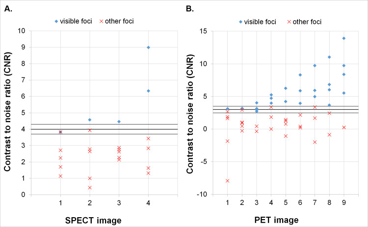
The data was acquired from the Jaszczak and NEMA phantoms. In the Jaszczak phantom Y-90 concentrations of 0.1 MBq/ml and 0.2 MBq/ml were used, while higher concentrations, up to 1.0 MBq/ml, were simulated by acquisition time extension with respect to the standard clinical protocol of 30 sec/projection for SPECT and 30 min/bed position for PET imaging. For NEMA phantom, the hot foci had concentrations of about 4 MB/ml and the background 0.1 or 0.0 MBq/ml. All of the acquired data was analysed both qualitatively and quantitatively. Qualitative assessment was conducted by six observers asked to identify the number of visible cold or hot foci. Inter-observer agreement was assessed. Quantitative analysis included calculations of contrast and contrast-to-noise ratio (CNR), and comparisons with the qualitative results.
For SPECT data up to two cold foci were discernible, while for PET four foci were visible. We have shown that CNR (with Rose criterion) is a good measure of foci visibility for both modalities. We also found good concordance of qualitative results for the Jaszczak phantom studies between the observers (corresponding Krippendorf's alpha coefficients of 0.76 to 0.84). In the NEMA phantom without background activity all foci were visible in SPECT/CT images. With isotope in the background, 5 of 6 spheres were discernible (CNR of 3.0 for the smallest foci). For PET studies all hot spheres were visible, regardless of the background activity.
PET Y-90 imaging provided better results than Bremsstrahlung based SPECT imaging. This indicates that PET/CT might become the method of choice in Y-90 post radioembolization imaging for visualisation of both necrotic and hot lesions in the liver.
本研究旨在评估 SPECT 和 PET Y-90 成像的可行性,并通过可视化不同同位素浓度下的冷热点来比较这两种模态。
数据来自 Jaszczak 和 NEMA 体模。在 Jaszczak 体模中,使用了 0.1 MBq/ml 和 0.2 MBq/ml 的 Y-90 浓度,而更高的浓度(高达 1.0 MBq/ml)则通过相对于 SPECT 的标准临床协议(30 秒/投影)和 PET 成像(30 分钟/床位位置)的采集时间延长来模拟。对于 NEMA 体模,热点的浓度约为 4 MB/ml,背景为 0.1 或 0.0 MBq/ml。所有采集的数据都进行了定性和定量分析。定性评估由六名观察者进行,要求他们识别可见的冷或热点的数量。评估了观察者间的一致性。定量分析包括对比度和对比噪声比(CNR)的计算,并与定性结果进行比较。
对于 SPECT 数据,最多可识别两个冷点,而对于 PET 则可识别四个焦点。我们表明,对于两种模态,CNR(用 Rose 标准)是焦点可见性的良好衡量标准。我们还发现,观察者之间对 Jaszczak 体模研究的定性结果具有很好的一致性(相应的 Krippendorf's alpha 系数为 0.76 至 0.84)。在没有背景活动的 NEMA 体模中,所有焦点在 SPECT/CT 图像中均可见。在有背景同位素的情况下,可识别出 6 个球体中的 5 个(最小焦点的 CNR 为 3.0)。对于 PET 研究,无论背景活动如何,所有热点均可见。
PET Y-90 成像的结果优于基于 Bremsstrahlung 的 SPECT 成像。这表明,对于 Y-90 后放射栓塞成像中的坏死和热点病变的可视化,PET/CT 可能成为首选方法。