IRCCS - Istituto di Ricerche Farmacologiche "Mario Negri", Centro Anna Maria Astori, Science and Technology Park Kilometro Rosso, Bergamo, Italy.
Unit of Nephrology and Dialysis, Azienda Socio Sanitaria Territoriale (ASST) Papa Giovanni XXIII, Bergamo, Italy.
Sci Rep. 2016 Jun 27;6:28445. doi: 10.1038/srep28445.
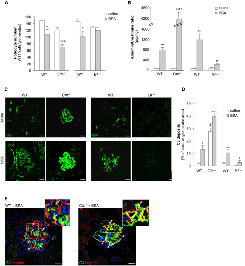
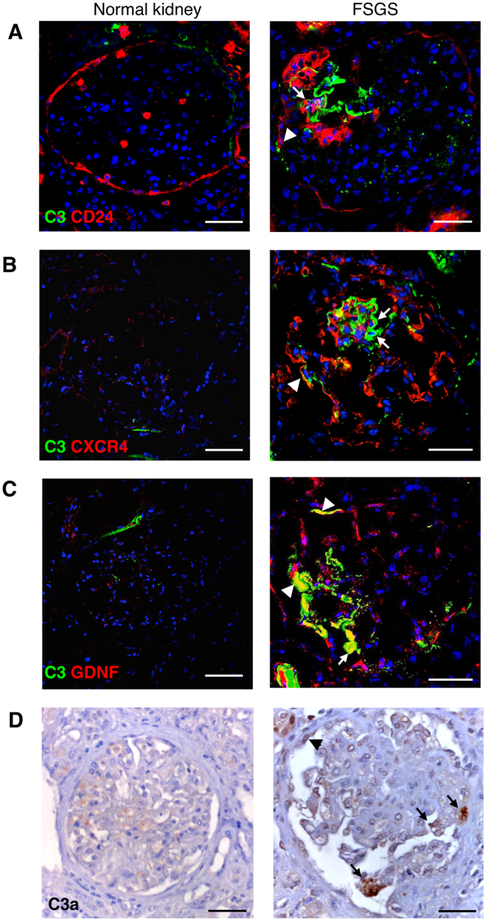
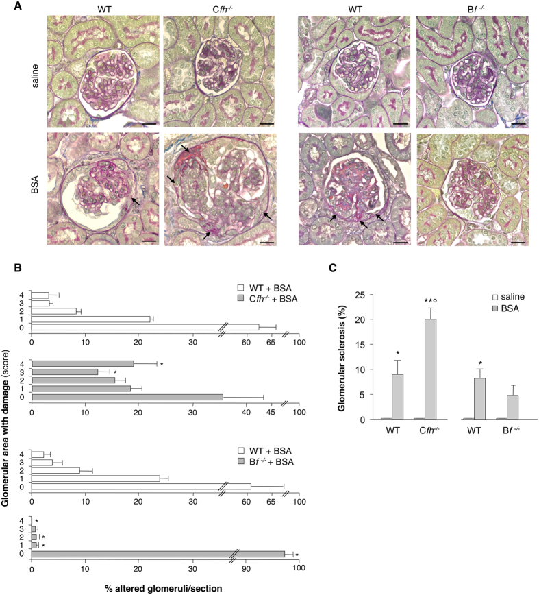
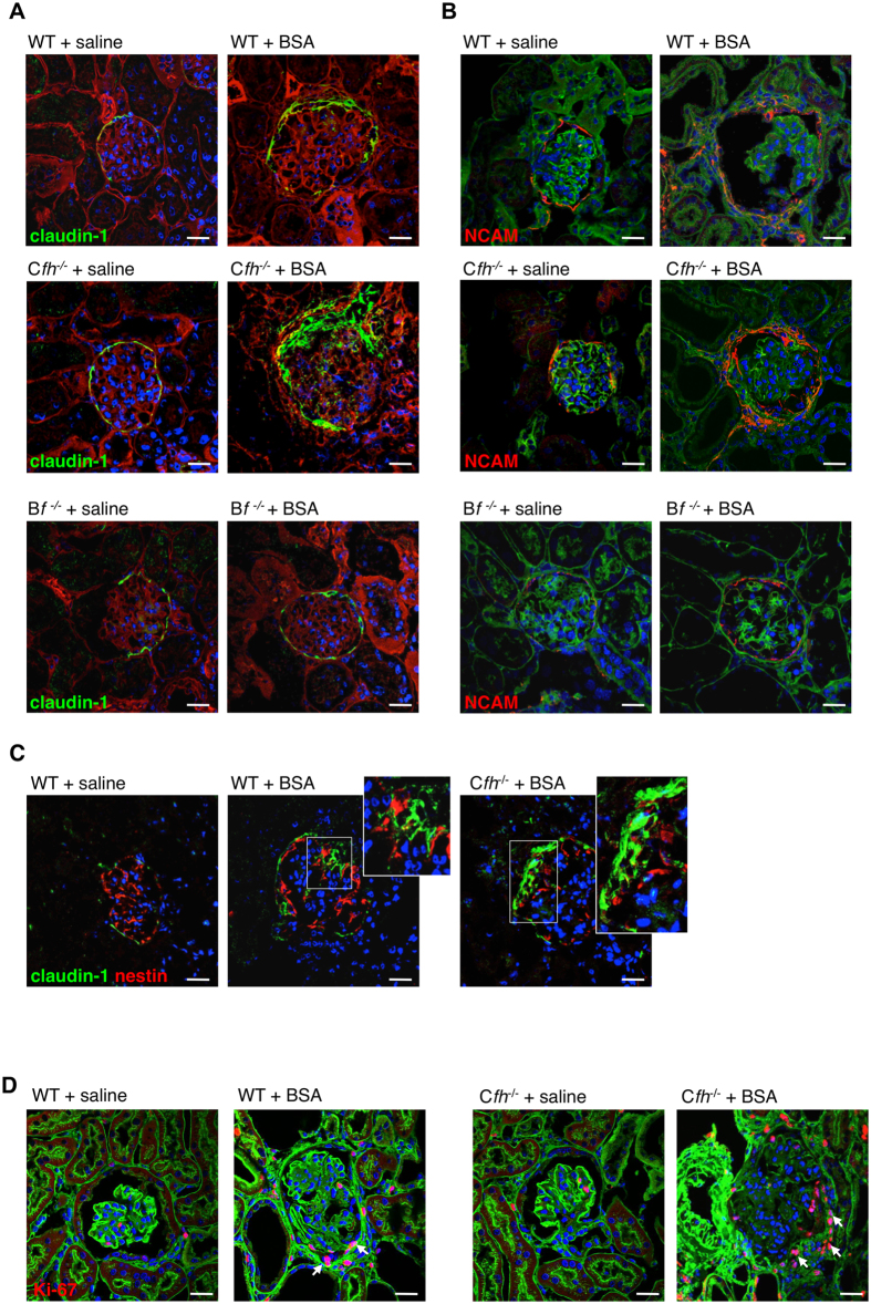
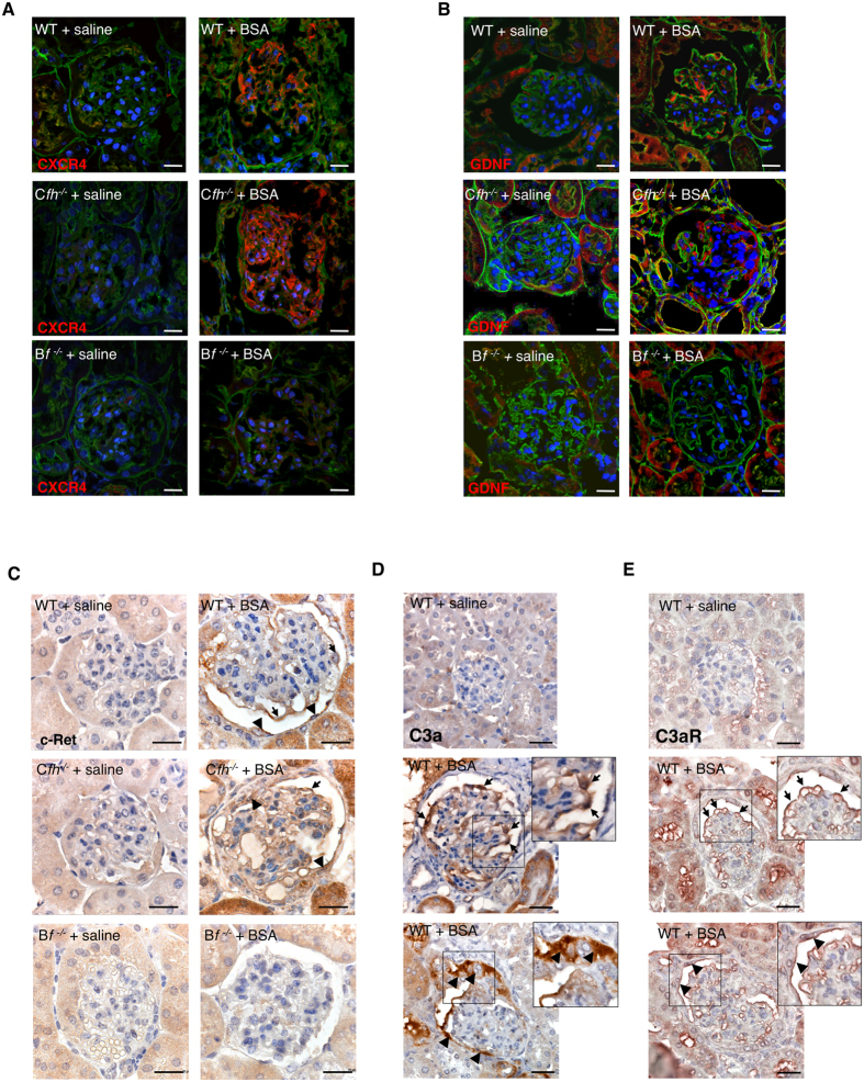
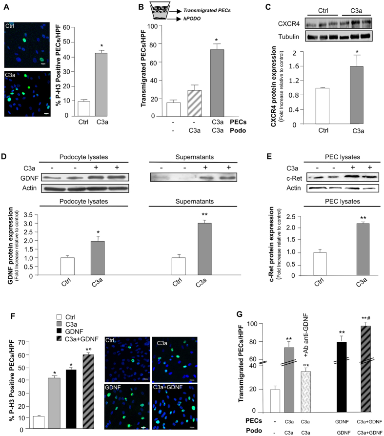
Podocyte loss is the initial event in the development of glomerulosclerosis, the structural hallmark of progressive proteinuric nephropathies. Understanding mechanisms underlying glomerular injury is the key challenge for identifying novel therapeutic targets. In mice with protein-overload induced by bovine serum albumin (BSA), we evaluated whether the alternative pathway (AP) of complement mediated podocyte depletion and podocyte-dependent parietal epithelial cell (PEC) activation causing glomerulosclerosis. Factor H (Cfh(-/-)) or factor B-deficient mice were studied in comparison with wild-type (WT) littermates. WT+BSA mice showed podocyte depletion accompanied by glomerular complement C3 and C3a deposits, PEC migration to capillary tuft, proliferation, and glomerulosclerosis. These changes were more prominent in Cfh(-/-) +BSA mice. The pathogenic role of AP was documented by data that factor B deficiency preserved glomerular integrity. In protein-overload mice, PEC dysregulation was associated with upregulation of CXCR4 and GDNF/c-Ret axis. In vitro studies provided additional evidence of a direct action of C3a on proliferation and CXCR4-related migration of PECs. These effects were enhanced by podocyte-derived GDNF. In patients with proteinuric nephropathy, glomerular C3/C3a paralleled PEC activation, CXCR4 and GDNF upregulation. These results indicate that mechanistically uncontrolled AP complement activation is not dispensable for podocyte-dependent PEC activation resulting in glomerulosclerosis.
足细胞丢失是肾小球硬化发展的初始事件,也是进行性蛋白尿性肾病的结构标志。理解肾小球损伤的机制是确定新的治疗靶点的关键挑战。在牛血清白蛋白(BSA)诱导的蛋白过载的小鼠中,我们评估了补体替代途径(AP)是否介导了足细胞耗竭和足细胞依赖性壁细胞(PEC)激活导致肾小球硬化。我们比较了补体因子 H(Cfh(-/-))或补体因子 B 缺陷型小鼠与野生型(WT)同窝仔鼠。WT+BSA 小鼠表现出足细胞耗竭,伴有肾小球补体 C3 和 C3a 沉积、PEC 迁移到毛细血管丛、增殖和肾小球硬化。在 Cfh(-/-) +BSA 小鼠中这些变化更为明显。补体因子 B 缺陷型保留了肾小球完整性,这证明了 AP 的致病作用。在蛋白过载小鼠中,PEC 失调与 CXCR4 和 GDNF/c-Ret 轴的上调有关。体外研究提供了更多证据表明 C3a 直接作用于 PEC 的增殖和 CXCR4 相关迁移。足细胞衍生的 GDNF 增强了这些作用。在蛋白尿性肾病患者中,肾小球 C3/C3a 与 PEC 激活、CXCR4 和 GDNF 上调平行。这些结果表明,机制上不受控制的 AP 补体激活对于导致肾小球硬化的足细胞依赖性 PEC 激活不是必需的。