Grandl Susanne, Sztrókay-Gaul Anikó, Mittone Alberto, Gasilov Sergey, Brun Emmanuel, Bravin Alberto, Mayr Doris, Auweter Sigrid D, Hellerhoff Karin, Reiser Maximilian, Coan Paola
Institute for Clinical Radiology, Ludwig-Maximilians-University Hospital, Munich, Germany.
Department of Physics, Ludwig-Maximilians-University, Garching, Germany.
PLoS One. 2016 Jun 30;11(6):e0158306. doi: 10.1371/journal.pone.0158306. eCollection 2016.
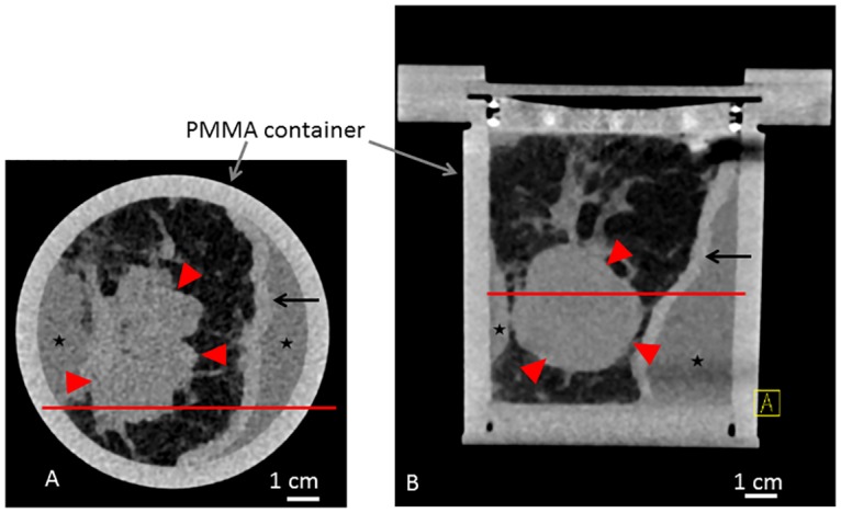
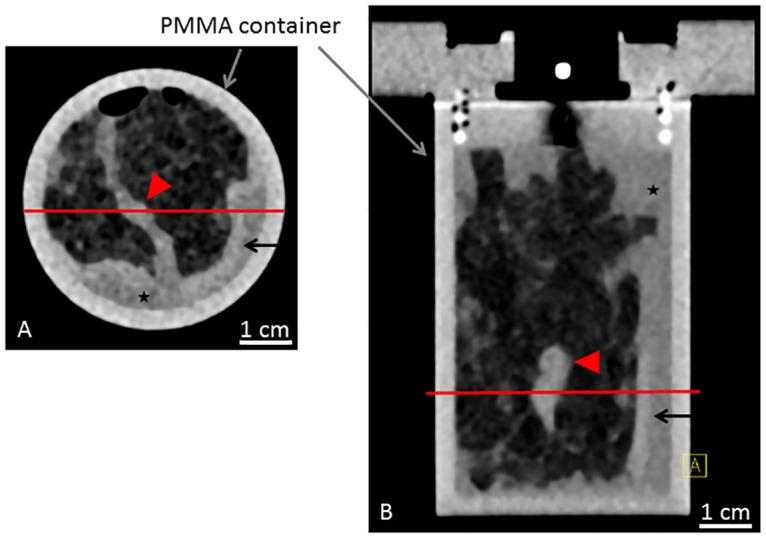
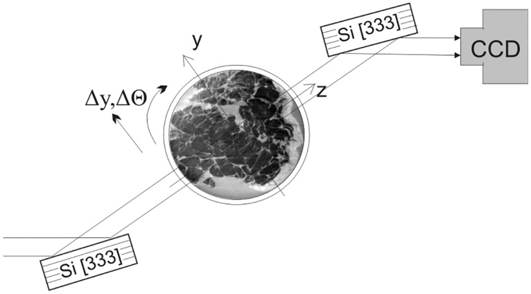
Neoadjuvant chemotherapy is the state-of-the-art treatment in advanced breast cancer. A correct visualization of the post-therapeutic tumor size is of high prognostic relevance. X-ray phase-contrast computed tomography (PC-CT) has been shown to provide improved soft-tissue contrast at a resolution formerly restricted to histopathology, at low doses. This study aimed at assessing ex-vivo the potential use of PC-CT for visualizing the effects of neoadjuvant chemotherapy on breast carcinoma.
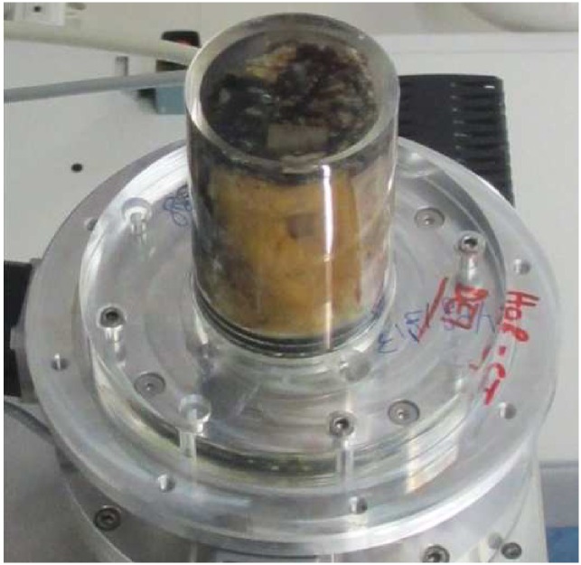
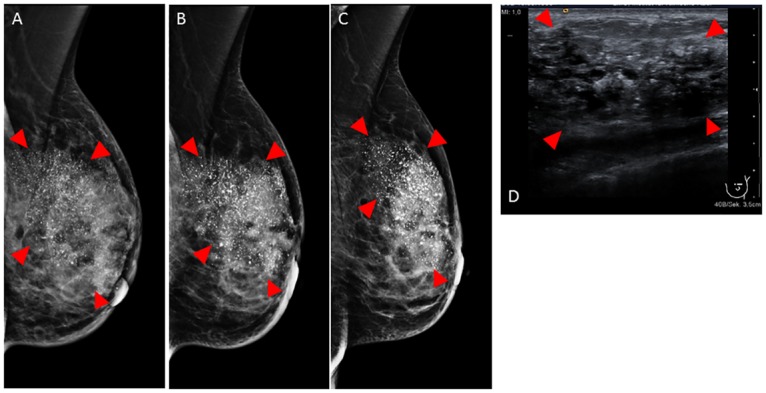
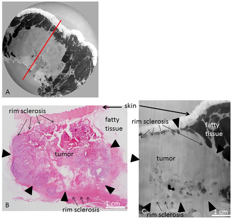
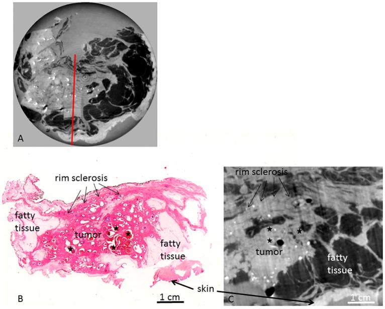
The analysis was performed on two ex-vivo formalin-fixed mastectomy samples containing an invasive carcinoma removed from two patients treated with neoadjuvant chemotherapy. Images were matched with corresponding histological slices. The visibility of typical post-therapeutic tissue changes was assessed and compared to results obtained with conventional clinical imaging modalities.
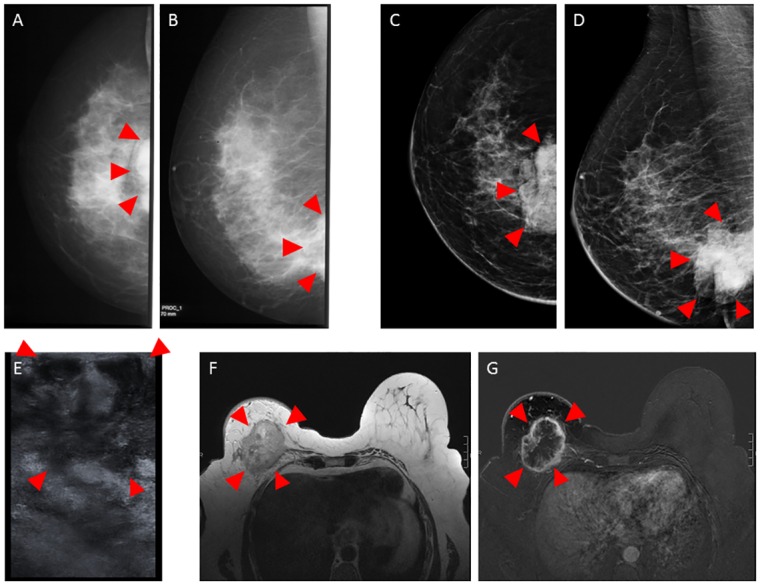
PC-CT depicted the different tissue types with an excellent correlation to histopathology. Post-therapeutic tissue changes were correctly visualized and the residual tumor mass could be detected. PC-CT outperformed clinical imaging modalities in the detection of chemotherapy-induced tissue alterations including post-therapeutic tumor size.
PC-CT might become a unique diagnostic tool in the prediction of tumor response to neoadjuvant chemotherapy. PC-CT might be used to assist during histopathological diagnosis, offering a high-resolution and high-contrast virtual histological tool for the accurate delineation of tumor boundaries.
新辅助化疗是晚期乳腺癌的先进治疗方法。准确显示治疗后肿瘤大小具有高度的预后相关性。X射线相衬计算机断层扫描(PC-CT)已被证明能够在低剂量下,以以前仅组织病理学所具备的分辨率提供更好的软组织对比度。本研究旨在评估PC-CT在体外对可视化新辅助化疗对乳腺癌的疗效的潜在用途。
对两个来自接受新辅助化疗的患者的离体福尔马林固定乳房切除样本进行分析,样本包含浸润性癌。将图像与相应的组织学切片进行匹配。评估典型治疗后组织变化的可见性,并与传统临床成像方式获得的结果进行比较。
PC-CT描绘的不同组织类型与组织病理学具有极好的相关性。治疗后组织变化得到正确显示,并且可以检测到残留肿瘤块。在检测化疗引起的组织改变(包括治疗后肿瘤大小)方面,PC-CT优于临床成像方式。
PC-CT可能成为预测肿瘤对新辅助化疗反应的独特诊断工具。PC-CT可用于在组织病理学诊断过程中提供辅助,为准确描绘肿瘤边界提供高分辨率和高对比度的虚拟组织学工具。