• 文献检索
  • 文档翻译
  • 深度研究
  • 学术资讯
  • Suppr Zotero 插件Zotero 插件
  • 邀请有礼
  • 套餐&价格
  • 历史记录
应用&插件
Suppr Zotero 插件Zotero 插件浏览器插件Mac 客户端Windows 客户端微信小程序
定价
高级版会员购买积分包购买API积分包
服务
文献检索文档翻译深度研究API 文档MCP 服务
关于我们
关于 Suppr公司介绍联系我们用户协议隐私条款
关注我们

Suppr 超能文献

核心技术专利:CN118964589B侵权必究
粤ICP备2023148730 号-1Suppr @ 2026

文献检索

告别复杂PubMed语法,用中文像聊天一样搜索,搜遍4000万医学文献。AI智能推荐,让科研检索更轻松。

立即免费搜索

文件翻译

保留排版,准确专业,支持PDF/Word/PPT等文件格式,支持 12+语言互译。

免费翻译文档

深度研究

AI帮你快速写综述,25分钟生成高质量综述,智能提取关键信息,辅助科研写作。

立即免费体验

使用手持式光学成像仪对人类乳腺癌进行非侵入性表面成像。

Noninvasive Surface Imaging of Breast Cancer in Humans using a Hand-held Optical Imager.

作者信息

Erickson-Bhatt Sarah J, Roman Manuela, Gonzalez Jean, Nunez Annie, Kiszonas Richard, Lopez-Penalver Cristina, Godavarty Anuradha

机构信息

Dept. of Biomedical Engineering, Florida International University, 10555 West Flagler St. EC2610, Miami, FL, USA 33174.

Dept. of Breast Radiology, Sylvester Comprehensive Cancer Center, 1475 N.W. 12th Ave., Miami, FL, USA 33136.

出版信息

Biomed Phys Eng Express. 2015 Dec;1(4). doi: 10.1088/2057-1976/1/4/045001. Epub 2015 Oct 23.

DOI:10.1088/2057-1976/1/4/045001
PMID:27366327
原文链接:https://pmc.ncbi.nlm.nih.gov/articles/PMC4922423/
Abstract

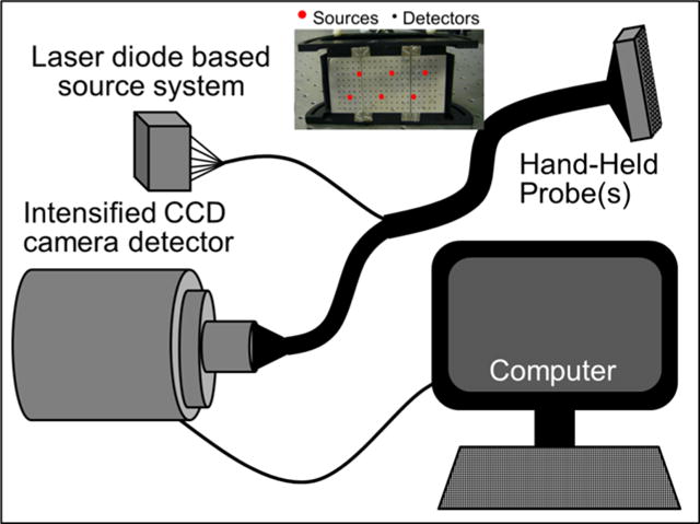
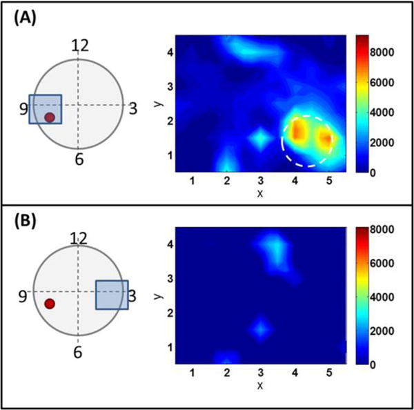
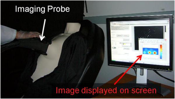
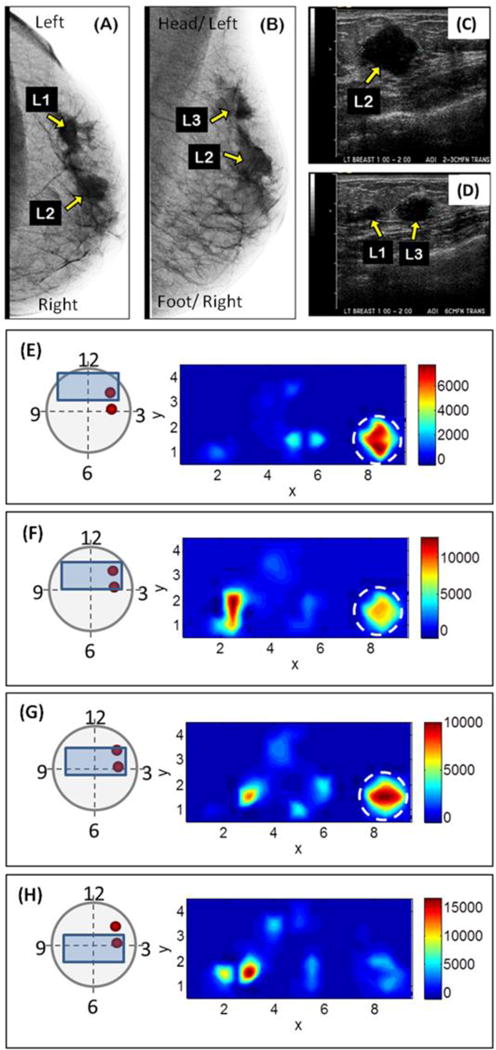
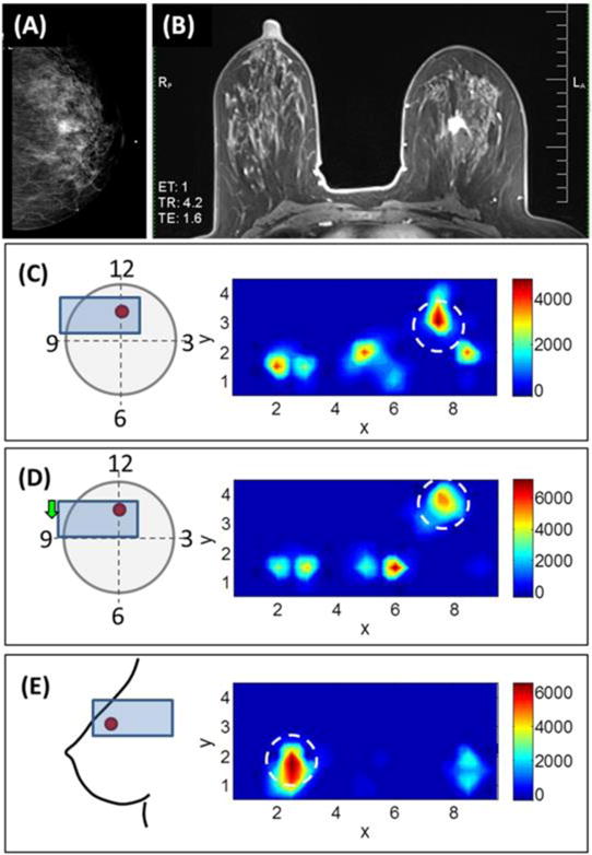
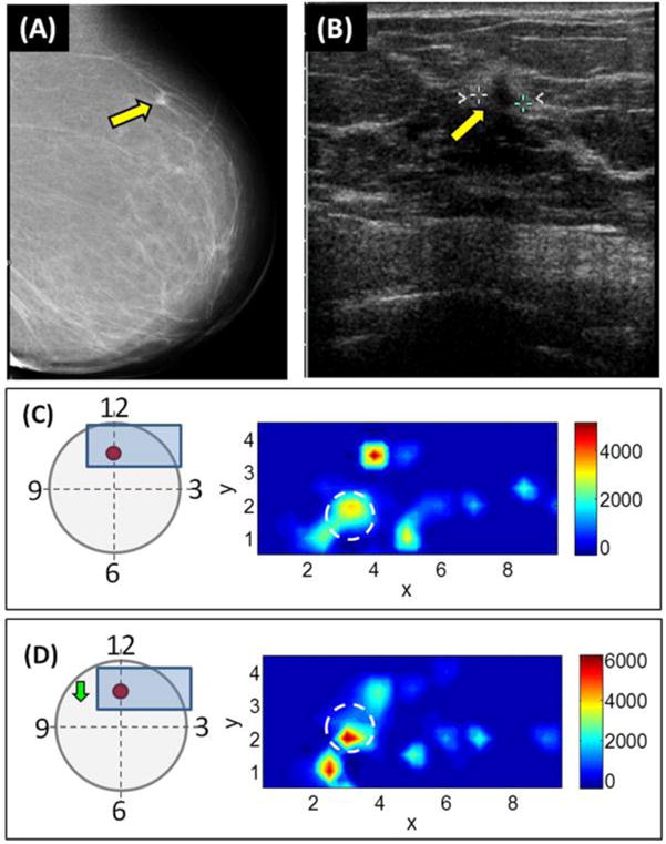
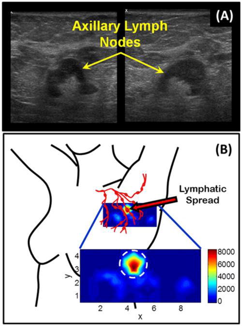
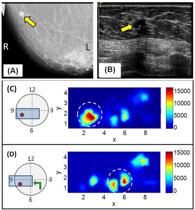
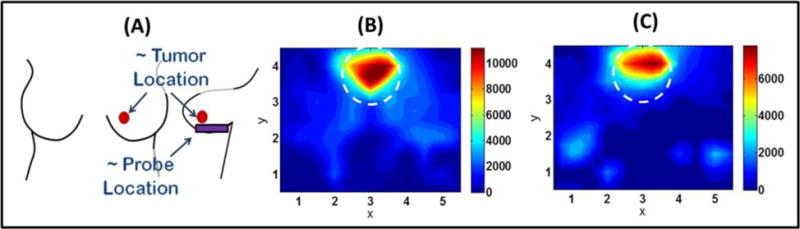
X-ray mammography, the current gold standard for breast cancer detection, has a 20% false-negative rate (cancer is undetected) and increases in younger women with denser breast tissue. Diffuse optical imaging (DOI) is a safe (nonionizing), and relatively inexpensive method for noninvasive imaging of breast cancer in human subjects (including dense breast tissues) by providing physiological information (e.g. oxy- and deoxy- hemoglobin concentration). At the Optical Imaging Laboratory, a hand-held optical imager has been developed which employs a breast contourable probe head to perform simultaneous illumination and detection of large surfaces towards near real-time imaging of human breast cancer. Gen-1 and gen-2 versions of the handheld optical imager have been developed and previously demonstrated imaging in tissue phantoms and healthy human subjects. Herein, the hand-held optical imagers are applied towards imaging of breast cancer subjects in an attempt to determine the ability of the imager to detect breast tumors. Five female human subjects (ages 51-74) diagnosed with breast cancer were imaged with the gen-1 optical imager prior to surgical intervention. One of the subjects was also imaged with the gen-2 optical imager. Both imagers use 785 nm laser diode sources and ICCD camera detectors to generate 2D surfaces maps of total hemoglobin absorption. The subjects lay in supine position and images were collected at various locations on both the ipsilateral (tumor-containing) and contralateral (non-tumor containing) breasts. The optical images (2D surface maps of optical absorption due to total hemoglobin concentration) show regions of higher intensity at the tumor location, which is indicative of increased vasculature and higher blood content due to the presence of the tumor. Additionally, a preliminary result indicates the potential to image lymphatic spread. This study demonstrates the potential of the hand-held optical devices to noninvasively image breast cancer in human subjects.

摘要

X线乳房摄影是目前乳腺癌检测的金标准,其假阴性率为20%(癌症未被检测到),并且在乳腺组织致密的年轻女性中该比例会更高。漫射光学成像(DOI)是一种安全(非电离)且相对廉价的方法,可通过提供生理信息(如氧合血红蛋白和脱氧血红蛋白浓度)对人体(包括致密乳腺组织)的乳腺癌进行无创成像。在光学成像实验室,已开发出一种手持式光学成像仪,它采用可贴合乳房的探头对大表面进行同时照明和检测,以实现对人类乳腺癌的近实时成像。已开发出第一代和第二代手持式光学成像仪,之前已在组织模型和健康人体受试者中展示了其成像效果。在此,将手持式光学成像仪应用于乳腺癌受试者的成像,以试图确定该成像仪检测乳腺肿瘤的能力。五名被诊断为乳腺癌的女性受试者(年龄51 - 74岁)在手术干预前用第一代光学成像仪进行了成像。其中一名受试者还用第二代光学成像仪进行了成像。两种成像仪均使用785 nm激光二极管光源和ICCD相机探测器来生成总血红蛋白吸收的二维表面图。受试者仰卧,在同侧(含肿瘤)和对侧(不含肿瘤)乳房的不同位置采集图像。光学图像(由于总血红蛋白浓度导致的光学吸收二维表面图)显示肿瘤位置处强度较高的区域,这表明由于肿瘤的存在,血管分布增加且血液含量更高。此外,初步结果表明有对淋巴扩散进行成像的潜力。这项研究证明了手持式光学设备对人体乳腺癌进行无创成像的潜力。

https://cdn.ncbi.nlm.nih.gov/pmc/blobs/0099/4922423/bed9c16673fe/nihms786754f9.jpg
https://cdn.ncbi.nlm.nih.gov/pmc/blobs/0099/4922423/00f1d97f19e1/nihms786754f1.jpg
https://cdn.ncbi.nlm.nih.gov/pmc/blobs/0099/4922423/c19d6fea95a8/nihms786754f2.jpg
https://cdn.ncbi.nlm.nih.gov/pmc/blobs/0099/4922423/60eb002c3b1f/nihms786754f3.jpg
https://cdn.ncbi.nlm.nih.gov/pmc/blobs/0099/4922423/1ee43276b52d/nihms786754f4.jpg
https://cdn.ncbi.nlm.nih.gov/pmc/blobs/0099/4922423/74d9b4ae6db5/nihms786754f5.jpg
https://cdn.ncbi.nlm.nih.gov/pmc/blobs/0099/4922423/581317fc5373/nihms786754f6.jpg
https://cdn.ncbi.nlm.nih.gov/pmc/blobs/0099/4922423/e04c0e445ef2/nihms786754f7.jpg
https://cdn.ncbi.nlm.nih.gov/pmc/blobs/0099/4922423/29c36f2e6689/nihms786754f8.jpg
https://cdn.ncbi.nlm.nih.gov/pmc/blobs/0099/4922423/bed9c16673fe/nihms786754f9.jpg

相似文献

1
Noninvasive Surface Imaging of Breast Cancer in Humans using a Hand-held Optical Imager.使用手持式光学成像仪对人类乳腺癌进行非侵入性表面成像。
Biomed Phys Eng Express. 2015 Dec;1(4). doi: 10.1088/2057-1976/1/4/045001. Epub 2015 Oct 23.
2
Hand-held optical imager (Gen-2): improved instrumentation and target detectability.手持式光学成像仪(Gen-2):改进的仪器设备和目标探测能力。
J Biomed Opt. 2012 Aug;17(8):081402-1. doi: 10.1117/1.JBO.17.8.081402.
3
Gen-2 hand-held optical imager towards cancer imaging: reflectance and transillumination phantom studies.用于癌症成像的 Gen-2 手持式光学成像仪:反射和透射幻影研究。
Sensors (Basel). 2012;12(2):1885-97. doi: 10.3390/s120201885. Epub 2012 Feb 10.
4
Three-dimensional fluorescence-enhanced optical tomography using a hand-held probe based imaging system.使用基于手持探头成像系统的三维荧光增强光学层析成像技术。
Med Phys. 2008 Jul;35(7):3354-63. doi: 10.1118/1.2940603.
5
In Vivo Observations of Rapid Scattered Light Changes Associated with Neurophysiological Activity与神经生理活动相关的快速散射光变化的体内观察
6
Automated coregistered imaging using a hand-held probe-based optical imager.使用基于手持探头的光学成像仪进行自动配准成像。
Rev Sci Instrum. 2010 Feb;81(2):023702. doi: 10.1063/1.3271019.
7
Three-dimensional fluorescence tomography of human breast tissues in vivo using a hand-held optical imager.利用手持式光学成像仪对人体乳腺组织进行三维荧光层析成像。
Phys Med Biol. 2013 Mar 7;58(5):1563-79. doi: 10.1088/0031-9155/58/5/1563. Epub 2013 Feb 15.
8
Optical imaging for breast cancer prescreening.乳腺癌筛查前的光学成像。
Breast Cancer (Dove Med Press). 2015 Jul 20;7:193-209. doi: 10.2147/BCTT.S51702. eCollection 2015.
9
In vivo absorption, scattering, and physiologic properties of 58 malignant breast tumors determined by broadband diffuse optical spectroscopy.通过宽带漫射光谱法测定58例乳腺恶性肿瘤的体内吸收、散射和生理特性。
J Biomed Opt. 2006 Jul-Aug;11(4):044005. doi: 10.1117/1.2337546.
10

引用本文的文献

1
Optimizing fresh specimen staining for rapid identification of tumor biomarkers during surgery.优化新鲜标本染色,以在手术期间快速鉴定肿瘤生物标志物。
Theranostics. 2017 Oct 17;7(19):4722-4734. doi: 10.7150/thno.21527. eCollection 2017.
2
Multispectral optoacoustic tomography of the human breast: characterisation of healthy tissue and malignant lesions using a hybrid ultrasound-optoacoustic approach.多光谱光声断层成像在人乳腺中的应用:使用混合超声-光声方法对健康组织和恶性病变的特征分析。
Eur Radiol. 2018 Feb;28(2):602-609. doi: 10.1007/s00330-017-5002-x. Epub 2017 Aug 7.
3
Heterodyne frequency-domain multispectral diffuse optical tomography of breast cancer in the parallel-plane transmission geometry.

本文引用的文献

1
Resolution of a Gen-2 handheld optical imager: diffuse and fluorescence imaging studies.第二代手持式光学成像仪的分辨率:漫射和荧光成像研究
Appl Opt. 2013 Nov 20;52(33):8060-6. doi: 10.1364/AO.52.008060.
2
Optical biomarkers for breast cancer derived from dynamic diffuse optical tomography.基于动态漫反射光学断层成像的乳腺癌光学生物标志物
J Biomed Opt. 2013 Sep;18(9):096012. doi: 10.1117/1.JBO.18.9.096012.
3
Monitoring the response to neoadjuvant hormone therapy for locally advanced breast cancer using three-dimensional time-resolved optical mammography.
平行平面透射几何结构下乳腺癌的外差频域多光谱扩散光学层析成像
Med Phys. 2016 Jul;43(7):4383. doi: 10.1118/1.4953830.
使用三维时分辨光乳腺成像监测局部晚期乳腺癌新辅助激素治疗的反应。
J Biomed Opt. 2013 May;18(5):56012. doi: 10.1117/1.JBO.18.5.056012.
4
Three-dimensional fluorescence tomography of human breast tissues in vivo using a hand-held optical imager.利用手持式光学成像仪对人体乳腺组织进行三维荧光层析成像。
Phys Med Biol. 2013 Mar 7;58(5):1563-79. doi: 10.1088/0031-9155/58/5/1563. Epub 2013 Feb 15.
5
Breast cancer: assessing response to neoadjuvant chemotherapy by using US-guided near-infrared tomography.乳腺癌:使用超声引导近红外层析成像评估新辅助化疗的反应。
Radiology. 2013 Feb;266(2):433-42. doi: 10.1148/radiol.12112415. Epub 2012 Dec 21.
6
Hand-held optical imager (Gen-2): improved instrumentation and target detectability.手持式光学成像仪(Gen-2):改进的仪器设备和目标探测能力。
J Biomed Opt. 2012 Aug;17(8):081402-1. doi: 10.1117/1.JBO.17.8.081402.
7
Baseline tumor oxygen saturation correlates with a pathologic complete response in breast cancer patients undergoing neoadjuvant chemotherapy.基线肿瘤氧饱和度与接受新辅助化疗的乳腺癌患者的病理完全缓解相关。
Cancer Res. 2012 Sep 1;72(17):4318-28. doi: 10.1158/0008-5472.CAN-12-0056. Epub 2012 Jul 9.
8
Gen-2 hand-held optical imager towards cancer imaging: reflectance and transillumination phantom studies.用于癌症成像的 Gen-2 手持式光学成像仪:反射和透射幻影研究。
Sensors (Basel). 2012;12(2):1885-97. doi: 10.3390/s120201885. Epub 2012 Feb 10.
9
Diffuse optical imaging and spectroscopy of the breast: a brief outline of history and perspectives.乳腺弥散光学成像和光谱学:历史与展望简述。
Photochem Photobiol Sci. 2012 Feb;11(2):241-50. doi: 10.1039/c1pp05230f. Epub 2011 Nov 18.
10
Diagnostic performance of a Near-Infrared Breast Imaging system as adjunct to mammography versus X-ray mammography alone.近红外乳腺成像系统与单独 X 射线乳腺摄影相比作为乳腺摄影的辅助诊断性能。
Eur Radiol. 2012 Feb;22(2):350-7. doi: 10.1007/s00330-011-2276-2. Epub 2011 Sep 27.