Department of Neurology, University of California, San Francisco, CA, United States; UCSF Comprehensive Epilepsy Center, United States.
Palo Alto University, Palo Alto, CA, United States.
Brain Lang. 2019 Jun;193:31-44. doi: 10.1016/j.bandl.2016.06.002. Epub 2016 Jul 5.
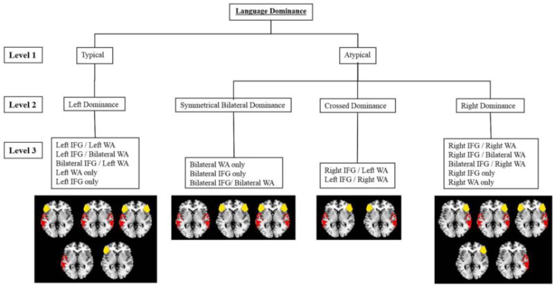
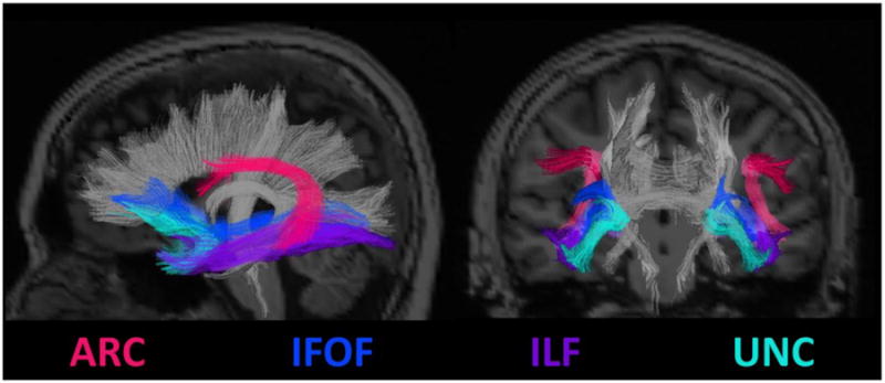
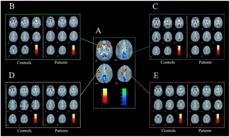
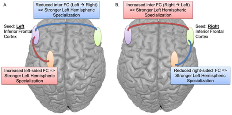
Advanced, noninvasive imaging has revolutionized our understanding of language networks in the brain and is reshaping our approach to the presurgical evaluation of patients with epilepsy. Functional magnetic resonance imaging (fMRI) has had the greatest impact, unveiling the complexity of language organization and reorganization in patients with epilepsy both pre- and postoperatively, while volumetric MRI and diffusion tensor imaging have led to a greater appreciation of structural and microstructural correlates of language dysfunction in different epilepsy syndromes. In this article, we review recent literature describing how unimodal and multimodal imaging has advanced our knowledge of language networks and their plasticity in epilepsy, with a focus on the most frequently studied epilepsy syndrome in adults, temporal lobe epilepsy (TLE). We also describe how new analytic techniques (i.e., graph theory) are leading to a refined characterization of abnormal brain connectivity, and how subject-specific imaging profiles combined with clinical data may enhance the prediction of both seizure and language outcomes following surgical interventions.
高级、非侵入性的成像技术已经彻底改变了我们对大脑语言网络的理解,并正在重塑我们对癫痫患者术前评估的方法。功能磁共振成像(fMRI)的影响最大,它揭示了癫痫患者手术前后语言组织和重组的复杂性,而容积磁共振成像和弥散张量成像则使我们更好地了解不同癫痫综合征中语言功能障碍的结构和微观结构相关性。在本文中,我们回顾了最近的文献,描述了单模态和多模态成像如何使我们对癫痫中的语言网络及其可塑性的认识得到了提高,重点介绍了成人中最常研究的癫痫综合征,即颞叶癫痫(TLE)。我们还描述了新的分析技术(例如,图论)如何导致对异常脑连接的精细描述,以及如何将个体特定的影像学特征与临床数据相结合,以提高对手术干预后癫痫发作和语言结果的预测。