Zhang Ying, Jia Xinling, Yang Jian, Li Qing, Yan Guofeng, Xu Zhongju, Wang Jingye
Department of Rehabilitation, Shanghai Xuhui Central Hospital, Shanghai Clinical Center, Chinese Academy of Sciences, No. 966, Huaihai Road, Shanghai 200031, China.
School of Life Sciences, Shanghai University, No. 99 Shangda Road, Shanghai 200444, China.
Biomed Res Int. 2016;2016:1859254. doi: 10.1155/2016/1859254. Epub 2016 Jun 20.
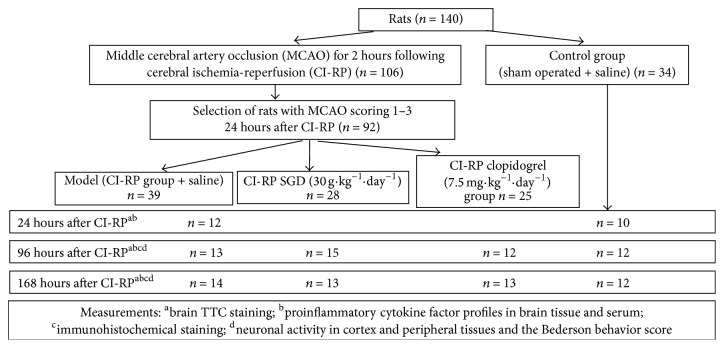
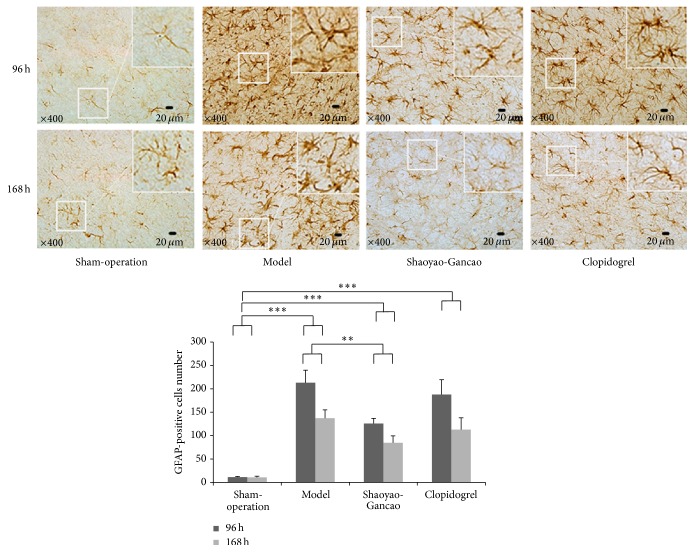
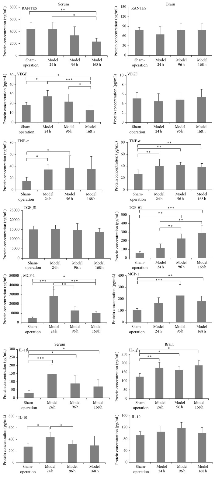
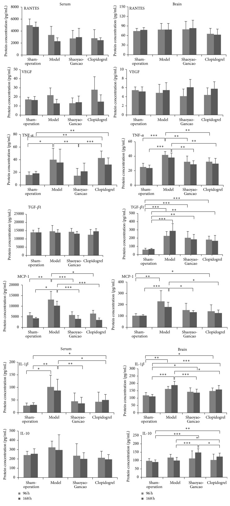
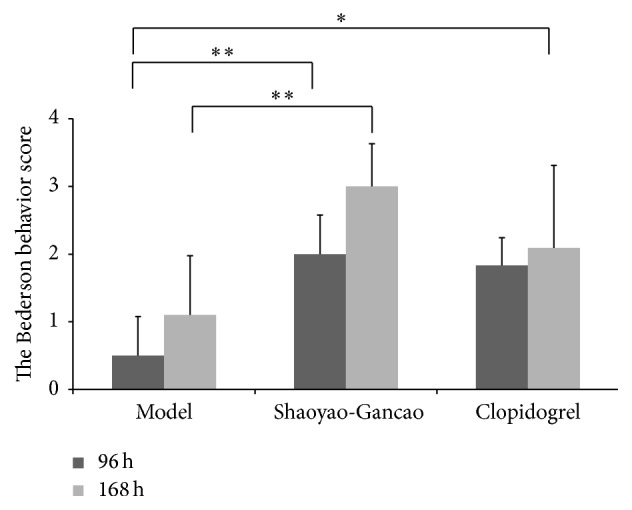
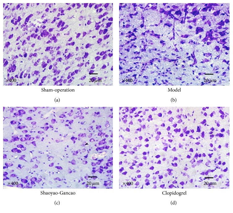
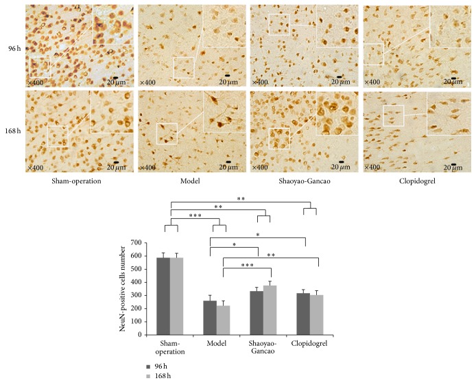
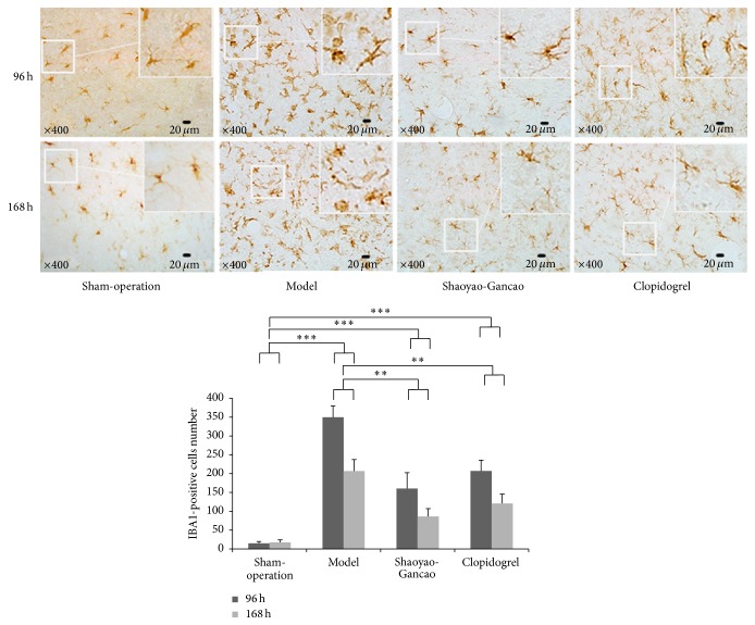
The mechanisms by which Shaoyao-Gancao decoction (SGD) inhibits the production of inflammatory cytokines in serum and brain tissue after cerebral ischemia-reperfusion (CI-RP) in rats were investigated. A right middle cerebral artery occlusion was used to induce CI-RP after which the rats were divided into model (n = 39), SGD (n = 28), clopidogrel (n = 25) and sham operated (n = 34) groups. The Bederson scale was used to evaluate changes in behavioral indices. The levels of IL-1β, TNF-α, MCP-1, IL-10, RANTES, VEGF, and TGF-β1 in the serum and infarcted brain tissues were measured. Nissl body and immunohistochemical staining methods were used to detect biochemical changes in neurons, microglial cells, and astrocytes. Serum levels of VEGF, TNF-α, MCP-1, IL-1β, and IL-10 increased significantly 24 h after CI-RP. In brain tissue, levels of TNF-α and IL-1β significantly increased 24 h after CI-RP, whereas levels of TGF-β1 and MCP-1 were significantly higher 96 h after CI-RP (P < 0.05). SGD or clopidogrel after CI-RP reduced TNF-α and IL-1β levels in brain tissue and serum levels of MCP-1, IL-1β, and IL-10. SGD increased the number of NeuN-positive cells in infarcted brain tissue and reduced the number of IBA1-positive and GFAP-positive cells. The efficacy of SGD was significantly higher than that of clopidogrel.
研究了芍药甘草汤(SGD)抑制大鼠脑缺血再灌注(CI-RP)后血清和脑组织中炎性细胞因子产生的机制。采用右侧大脑中动脉闭塞法诱导CI-RP,然后将大鼠分为模型组(n = 39)、SGD组(n = 28)、氯吡格雷组(n = 25)和假手术组(n = 34)。采用Bederson量表评估行为指标的变化。检测血清和梗死脑组织中IL-1β、TNF-α、MCP-1、IL-10、RANTES、VEGF和TGF-β1的水平。采用尼氏小体和免疫组织化学染色方法检测神经元、小胶质细胞和星形胶质细胞的生化变化。CI-RP后24小时,血清VEGF、TNF-α、MCP-1、IL-1β和IL-10水平显著升高。在脑组织中,CI-RP后24小时TNF-α和IL-1β水平显著升高,而CI-RP后96小时TGF-β1和MCP-1水平显著升高(P < 0.05)。CI-RP后给予SGD或氯吡格雷可降低脑组织中TNF-α和IL-1β水平以及血清中MCP-1、IL-1β和IL-10水平。SGD增加了梗死脑组织中NeuN阳性细胞的数量,减少了IBA1阳性和GFAP阳性细胞的数量。SGD的疗效显著高于氯吡格雷。