Lee Jun-Hyung, Hyun Jin-Soo, Kang Da-Yeong, Lee Hee-Jeong, Park Sang-Gon
Department of Internal Medicine, Chosun University Hospital, 365 Pilmun-daero, Dong-gu, Gwangju, 501-717, Republic of Korea.
Department of Internal Medicine, Hemato-Oncology, Chosun University Hospital, 365 Pilmun-daero, Dong-gu, Gwangju, 501-717, Republic of Korea.
J Med Case Rep. 2016 Jul 16;10:195. doi: 10.1186/s13256-016-0991-7.
Mucormycosis is a rare and life-threatening invasive fungal infection. Pulmonary mucormycosis commonly occurs in patients with severe neutropenia. Typically, pulmonary mucormycosis causes tissue necrosis resulting from angioinvasion and subsequent thrombosis, so most cases can occur with necrotizing pneumonia and/or hemoptysis. Some complex cases may invade adjacent organs, such as the mediastinum, pericardium, and chest wall. However, to the best our knowledge there is little known regarding bronchoesophageal fistula due to pulmonary mucormycosis after induction chemotherapy for acute myeloid leukemia. We present a case report about this unusual presentation.
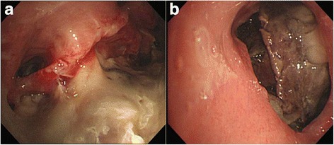
A 51-year-old Korean man was diagnosed as having acute myeloid leukemia and received induction chemotherapy. After prolonged severe neutropenia, he complained of coughing with aspiration. Imaging showed a bronchoesophageal fistula with extensive necrotizing pneumonia in the middle and lower lobes of his right lung. Bronchoscopy showed near total tissue necrosis in the middle lobe of his right lung, creating an orifice. A bronchial scope was passed through and was able to be connected with his esophagus; a bronchial wall biopsy was performed. Esophagoscopy revealed a large linear defect of his esophageal wall 30 cm from the incision that may have connected with the bronchus. A bronchial biopsy showed typical hyphae with necrotic tissue, indicating pulmonary mucormycosis. He was given amphotericin B, and a wide excision of lung and esophagus was planned. However, he suddenly died due to massive hemoptysis.
Here we present an extremely rare case of bronchoesophageal fistula with severe necrotizing pneumonia due to pulmonary mucormycosis.
毛霉菌病是一种罕见且危及生命的侵袭性真菌感染。肺毛霉菌病常见于严重中性粒细胞减少的患者。通常,肺毛霉菌病会因血管侵袭和随后的血栓形成导致组织坏死,因此大多数病例会伴有坏死性肺炎和/或咯血。一些复杂病例可能会侵犯相邻器官,如纵隔、心包和胸壁。然而,据我们所知,关于急性髓系白血病诱导化疗后肺毛霉菌病导致支气管食管瘘的情况知之甚少。我们报告了一例这种不寻常表现的病例。
一名51岁的韩国男性被诊断为急性髓系白血病并接受了诱导化疗。在长期严重中性粒细胞减少后,他主诉咳嗽伴误吸。影像学检查显示右肺中叶和下叶有支气管食管瘘伴广泛坏死性肺炎。支气管镜检查显示右肺中叶几乎完全组织坏死,形成一个孔口。将支气管镜通过该孔口并能够与他的食管相连;进行了支气管壁活检。食管镜检查显示距切口30 cm处食管壁有一个大的线性缺损,可能与支气管相连。支气管活检显示典型的菌丝和坏死组织,提示肺毛霉菌病。给予他两性霉素B治疗,并计划进行肺和食管的广泛切除。然而,他因大量咯血突然死亡。
我们在此报告一例极其罕见的因肺毛霉菌病导致支气管食管瘘伴严重坏死性肺炎的病例。