Zamboglou Constantinos, Schiller Florian, Fechter Tobias, Wieser Gesche, Jilg Cordula Annette, Chirindel Alin, Salman Nasr, Drendel Vanessa, Werner Martin, Mix Michael, Meyer Philipp Tobias, Grosu Anca Ligia
Department of Radiation Oncology, University Medical Center Freiburg, Germany; German Cancer Consortium (DKTK), Partner Site Freiburg, Germany;
Department of Nuclear Medicine, University Medical Center Freiburg, Germany; German Cancer Consortium (DKTK), Partner Site Freiburg, Germany;
Theranostics. 2016 Jun 18;6(10):1619-28. doi: 10.7150/thno.15344. eCollection 2016.
We performed a voxel-wise comparison of (68)Ga-HBED-CC-PSMA PET/CT with prostate histopathology to evaluate the performance of (68)Ga-HBED-CC-PSMA for the detection and delineation of primary prostate cancer (PCa).
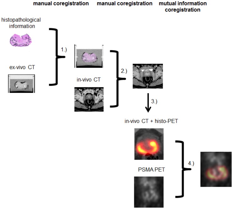
Nine patients with histopathological proven primary PCa underwent (68)Ga-HBED-CC-PSMA PET/CT followed by radical prostatectomy. Resected prostates were scanned by ex-vivo CT in a special localizer and histopathologically prepared. Histopathological information was matched to ex-vivo CT. PCa volume (PCa-histo) and non-PCa tissue in the prostate (NPCa-histo) were processed to obtain a PCa-model, which was adjusted to PET-resolution (histo-PET). Each histo-PET was coregistered to in-vivo PSMA-PET/CT data.
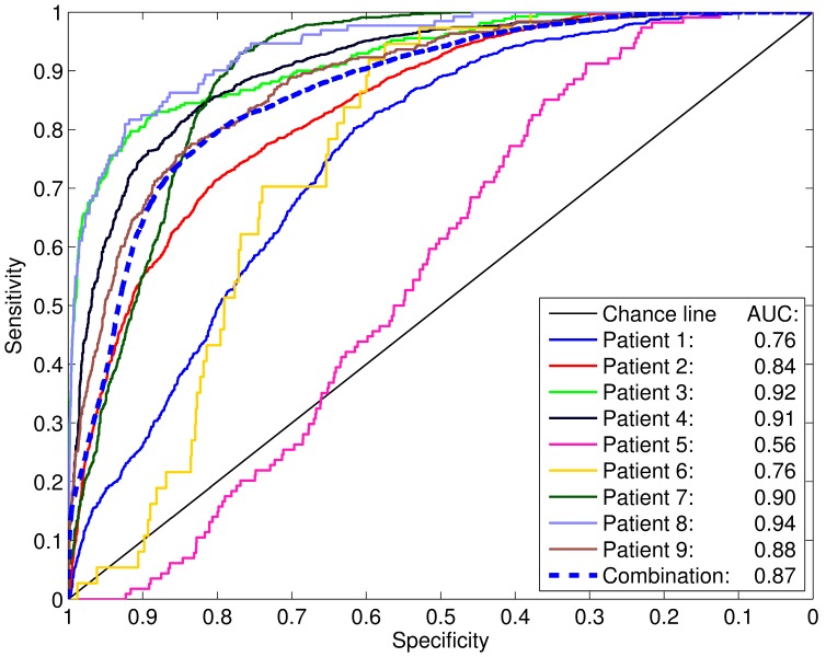
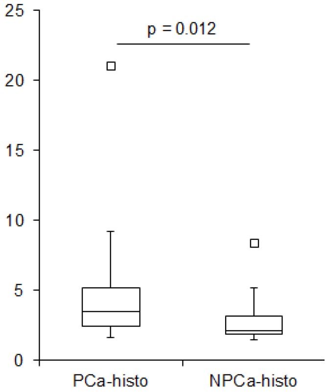
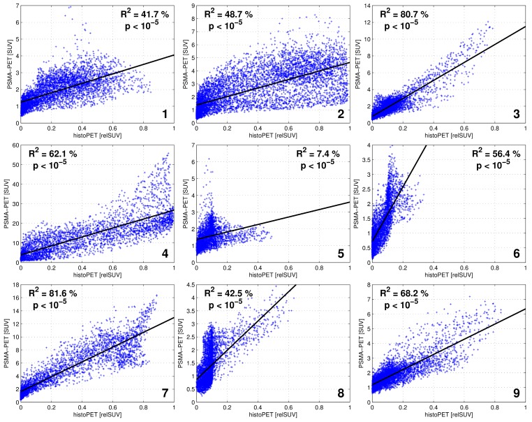
Analysis of spatial overlap between histo-PET and PSMA PET revealed highly significant correlations (p < 10(-5)) in nine patients and moderate to high coefficients of determination (R²) from 42 to 82 % with an average of 60 ± 14 % in eight patients (in one patient R(2) = 7 %). Mean SUVmean in PCa-histo and NPCa-histo was 5.6 ± 6.1 and 3.3 ± 2.5 (p = 0.012). Voxel-wise receiver-operating characteristic (ROC) analyses comparing the prediction by PSMA-PET with the non-smoothed tumor distribution from histopathology yielded an average area under the curve of 0.83 ± 0.12. Absolute and relative SUV (normalized to SUVmax) thresholds for achieving at least 90 % sensitivity were 3.19 ± 3.35 and 0.28 ± 0.09, respectively.
Voxel-wise analyses revealed good correlations of (68)Ga-HBED-CC-PSMA PET/CT and histopathology in eight out of nine patients. Thus, PSMA-PET allows a reliable detection and delineation of PCa as basis for PET-guided focal therapies.
我们对(68)Ga-HBED-CC-PSMA PET/CT与前列腺组织病理学进行了体素级比较,以评估(68)Ga-HBED-CC-PSMA在检测和勾勒原发性前列腺癌(PCa)方面的性能。
9例经组织病理学证实为原发性PCa的患者接受了(68)Ga-HBED-CC-PSMA PET/CT检查,随后进行了根治性前列腺切除术。切除的前列腺在特殊定位器中进行离体CT扫描,并进行组织病理学处理。将组织病理学信息与离体CT进行匹配。对前列腺中的PCa体积(PCa-histo)和非PCa组织(NPCa-histo)进行处理,以获得PCa模型,并将其调整为PET分辨率(histo-PET)。将每个histo-PET与体内PSMA-PET/CT数据进行配准。
对histo-PET和PSMA PET之间的空间重叠分析显示,9例患者具有高度显著的相关性(p < 10(-5)),8例患者的决定系数(R²)为中等至高,范围为42%至82%,平均为60 ± 14%(其中一例患者R(2) = 7%)。PCa-histo和NPCa-histo中的平均SUVmean分别为5.6 ± 6.1和3.3 ± 2.5(p = 0.012)。将PSMA-PET的预测与组织病理学中未平滑的肿瘤分布进行比较的体素级接受者操作特征(ROC)分析得出,曲线下平均面积为0.83 ± 0.12。达到至少90%敏感性的绝对和相对SUV(归一化至SUVmax)阈值分别为3.19 ± 3.35和0.28 ± 0.09。
体素级分析显示,9例患者中有8例(68)Ga-HBED-CC-PSMA PET/CT与组织病理学具有良好的相关性。因此,PSMA-PET能够可靠地检测和勾勒PCa,作为PET引导下局部治疗的基础。