Hettich Michael, Braun Friederike, Bartholomä Mark D, Schirmbeck Reinhold, Niedermann Gabriele
1. Department of Radiation Oncology, Medical Center - University of Freiburg, D-79106 Freiburg, Germany.; 3. Faculty of Biology, University of Freiburg, D-79104 Freiburg, Germany.
2. Department of Nuclear Medicine, Medical Center - University of Freiburg, D-79106 Freiburg, Germany.; 3. Faculty of Biology, University of Freiburg, D-79104 Freiburg, Germany.
Theranostics. 2016 Jun 18;6(10):1629-40. doi: 10.7150/thno.15253. eCollection 2016.
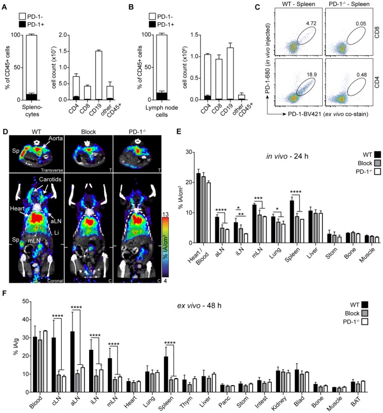
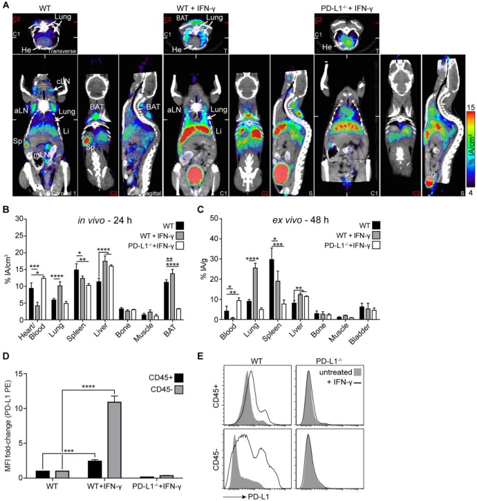
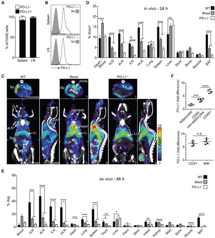
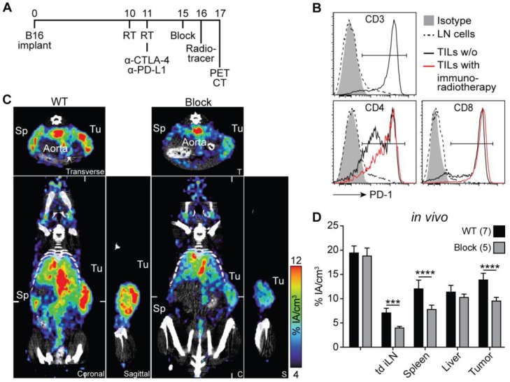
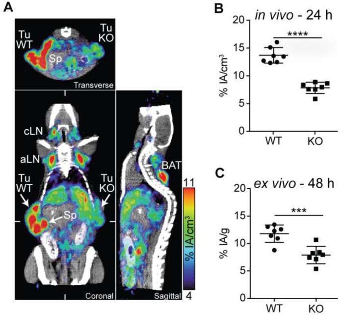
Checkpoint-blocking antibodies like those targeting the PD-1/PD-L1 pathway have revolutionized oncology. We developed radiotracers based on therapeutic checkpoint-blocking antibodies permitting sensitive and high-resolution PET imaging of both PD-1 and PD-L1 in immunocompetent mice. ImmunoPET of naive mice revealed similar overall expression patterns for PD-1 and PD-L1 in secondary lymphoid organs (spleen and lymph nodes). Interestingly, PD-L1 was also detected in brown adipose tissue (BAT), confirming the notion that BAT is immunologically relevant. Under pathophysiological conditions, strong expression of the receptor/ligand pair was also found in non-lymphoid tissues. Both were specifically detected in malignant tumors. PD-1 was readily detected after combined immunoradiotherapy causing massive tumor infiltration by PD-1+ lymphocytes. PD-L1 tracer uptake was reduced in PD-L1 knockout tumors. Moreover, monitoring the expression changes of PD-L1 in response to its main inducer, the effector T cell cytokine IFN-γ, revealed robust upregulation in the lung. This suggests that T cell responses in the lung, a vital organ continuously exposed to a variety of antigens, are strongly restrained by the PD-1 checkpoint. In turn, this could explain the association of PD-1 checkpoint inhibition with potentially fatal immune-mediated pneumonitis and partially also its efficacy in lung cancer.
像那些靶向PD-1/PD-L1通路的检查点阻断抗体已经彻底改变了肿瘤学。我们基于治疗性检查点阻断抗体开发了放射性示踪剂,能够在免疫健全的小鼠体内对PD-1和PD-L1进行灵敏且高分辨率的PET成像。对未接触过抗原的小鼠进行免疫PET成像显示,在二级淋巴器官(脾脏和淋巴结)中,PD-1和PD-L1的整体表达模式相似。有趣的是,在棕色脂肪组织(BAT)中也检测到了PD-L1,这证实了棕色脂肪组织具有免疫相关性这一观点。在病理生理条件下,在非淋巴组织中也发现了受体/配体对的强烈表达。两者在恶性肿瘤中均被特异性检测到特异性检测到。在联合免疫放疗导致大量PD-1+淋巴细胞浸润肿瘤后,很容易检测到PD-1。在PD-L1基因敲除肿瘤中,PD-L1示踪剂摄取减少。此外,监测PD-L1对其主要诱导物效应T细胞细胞因子IFN-γ的表达变化,发现肺中有强烈的上调。这表明,在不断接触各种抗原的重要器官肺中,T细胞反应受到PD-1检查点的强烈抑制。反过来,这可以解释PD-1检查点抑制与潜在致命的免疫介导性肺炎之间的关联,也部分解释了其在肺癌中的疗效。