Mądry Wojciech, Karolczak Maciej Aleksander
Department of Cardiac and General Pediatric Surgery, Warsaw Medical University Independent Public Paediatric Clinical Hospital in Warsaw, Poland.
J Ultrason. 2016 Jun;16(65):135-44. doi: 10.15557/JoU.2016.0015. Epub 2016 Jun 29.
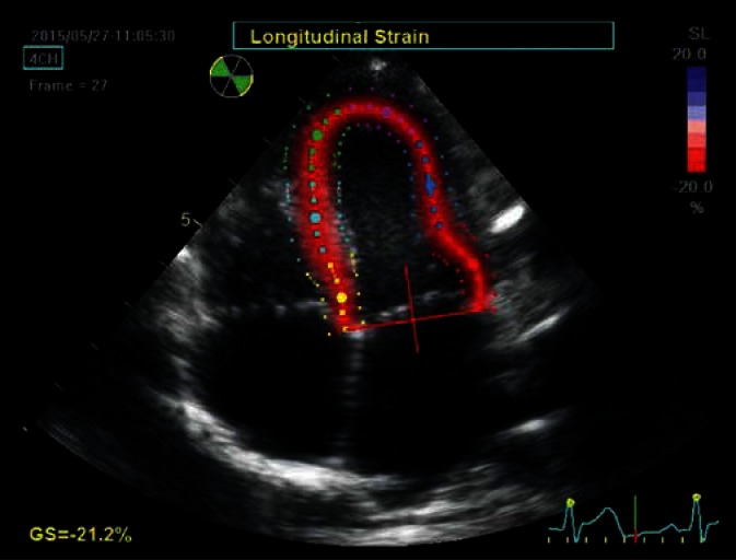
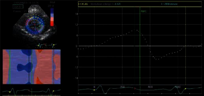
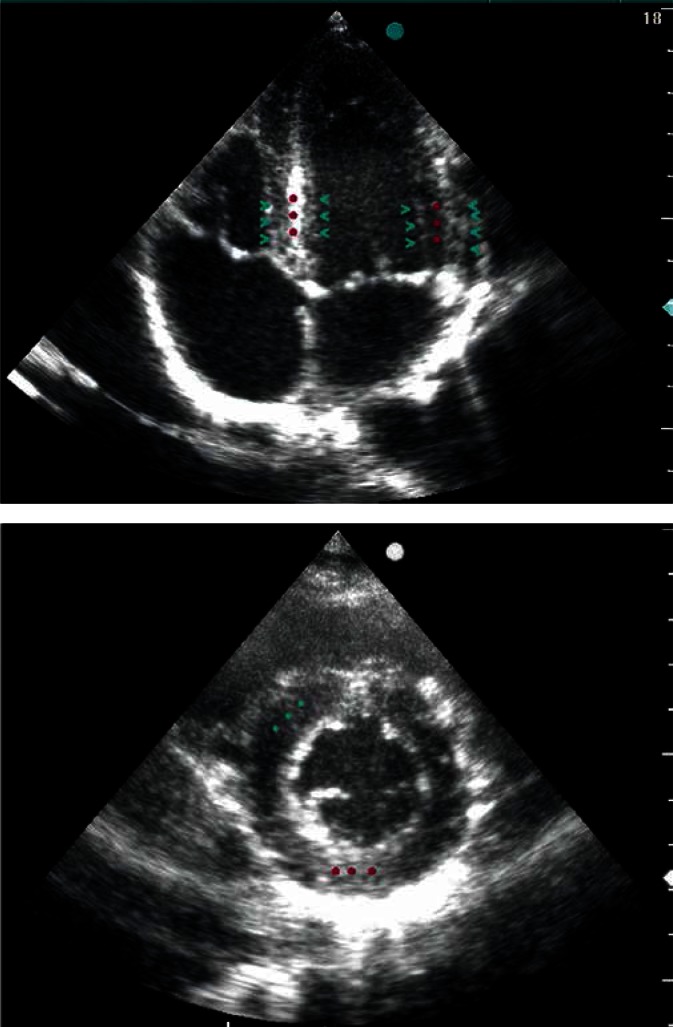
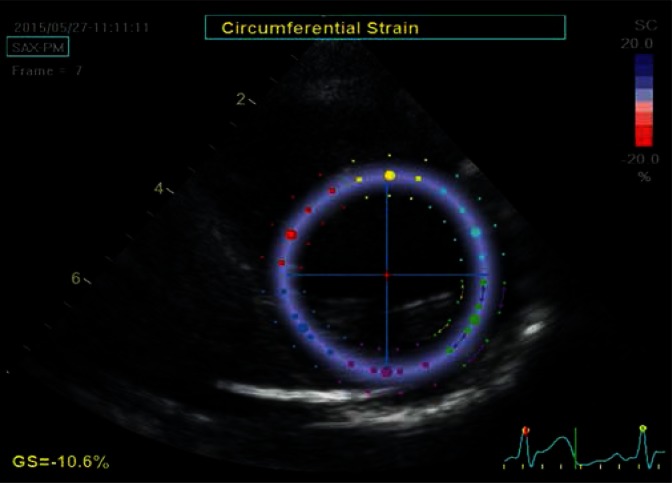
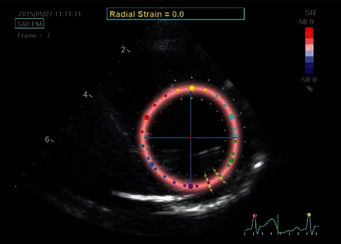
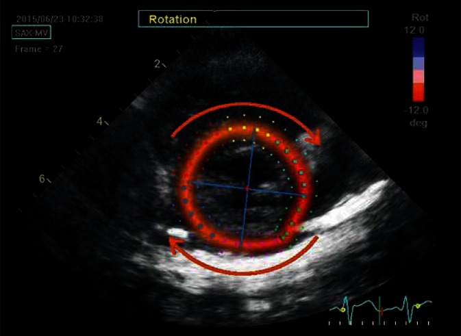
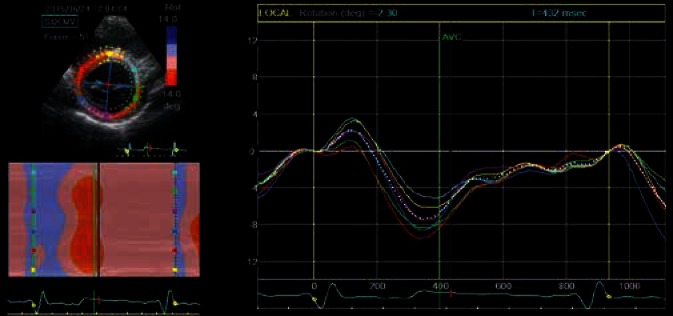
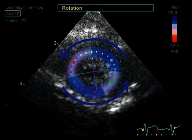
In this paper, the authors attempt to concisely present the anatomical and pathophysiological bases as well as the principles for echocardiographic evaluation of mechanical aspects of cardiac function based on speckle tracking method. This technique uses a phenomenon involving the formation of characteristic image units, referred to as speckles or acoustic markers, which are stable during cardiac cycle, on a two-dimensional echocardiographic picture. Changes in the position of these speckles throughout the cardiac cycle, which are monitored and analyzed semi-automatically by a computer system, reflect deformation of both, cardiac ventricle as a whole as well as its individual anatomical segments. The values of strain and the strain rate, as well as the range and velocity of the movement of these markers, which are in close relationship with multiple hemodynamic parameters, can be visualized as various types of charts - linear, two- and three-dimensional - as well as numerical values, enabling deeper insight into the mechanical and hemodynamic aspects of cardiac function in health and disease. The use of information obtained based on speckle tracking echocardiography allows to understand previously unclear mechanisms of physiological and pathophysiological processes. The first part of the study discusses the formation of a two-dimensional ultrasound image and the speckles, as well as the technical aspects of tracking their movement. The second part presents in more detail the methodology of speckle-tracking echocardiography, the characteristic abnormalities of cardiac mechanics presenting in different clinical entities, and the limitations related to given clinical and technical issues.
在本文中,作者试图简要介绍基于散斑追踪法的心脏功能力学方面的解剖学和病理生理学基础以及超声心动图评估原则。该技术利用了一种现象,即在二维超声心动图图像上形成特征性图像单元,称为散斑或声学标记,它们在心动周期中是稳定的。这些散斑在整个心动周期中的位置变化由计算机系统进行半自动监测和分析,反映了整个心室及其各个解剖节段的变形。应变和应变率的值,以及这些标记的移动范围和速度,与多个血流动力学参数密切相关,可以以各种类型的图表(线性、二维和三维)以及数值的形式可视化,从而能够更深入地了解健康和疾病状态下心脏功能的力学和血流动力学方面。基于散斑追踪超声心动图获得的信息的使用有助于理解以前不清楚的生理和病理生理过程机制。研究的第一部分讨论了二维超声图像和散斑的形成,以及追踪它们移动的技术方面。第二部分更详细地介绍了散斑追踪超声心动图的方法、不同临床实体中出现的心脏力学特征异常以及与特定临床和技术问题相关的局限性。