Radiology, Mayo Clinic, Rochester, Minnesota, USA.
Cardiovascular Diseases, Mayo Clinic, Rochester, Minnesota, USA.
J Magn Reson Imaging. 2017 Nov;46(5):1361-1367. doi: 10.1002/jmri.25678. Epub 2017 Feb 25.
To evaluate if cardiac magnetic resonance elastography (MRE) can measure increased stiffness in patients with cardiac amyloidosis. Myocardial tissue stiffness plays an important role in cardiac function. A noninvasive quantitative imaging technique capable of measuring myocardial stiffness could aid in disease diagnosis, therapy monitoring, and disease prognostic strategies. We recently developed a high-frequency cardiac MRE technique capable of making noninvasive stiffness measurements.
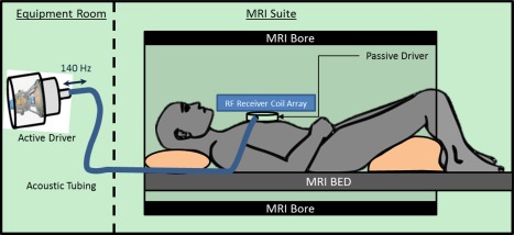
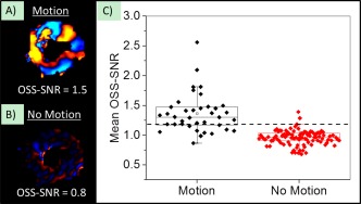
In all, 16 volunteers and 22 patients with cardiac amyloidosis were enrolled in this study after Institutional Review Board approval and obtaining formal written consent. All subjects were imaged head-first in the supine position in a 1.5T closed-bore MR imager. 3D MRE was performed using 5 mm isotropic resolution oblique short-axis slices and a vibration frequency of 140 Hz to obtain global quantitative in vivo left ventricular stiffness measurements. The median stiffness was compared between the two cohorts. An octahedral shear strain signal-to-noise ratio (OSS-SNR) threshold of 1.17 was used to exclude exams with insufficient motion amplitude.
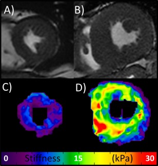
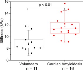
Five volunteers and six patients had to be excluded from the study because they fell below the 1.17 OSS-SNR threshold. The myocardial stiffness of cardiac amyloid patients (median: 11.4 kPa, min: 9.2, max: 15.7) was significantly higher (P = 0.0008) than normal controls (median: 8.2 kPa, min: 7.2, max: 11.8).
This study demonstrates the feasibility of 3D high-frequency cardiac MRE as a contrast-agent-free diagnostic imaging technique for cardiac amyloidosis.
2 Technical Efficacy: Stage 2 J. Magn. Reson. Imaging 2017;46:1361-1367.
评估心脏磁共振弹性成像(MRE)是否可测量心脏淀粉样变患者的僵硬度增加。心肌组织的僵硬度在心功能中起着重要作用。一种能够测量心肌僵硬度的非侵入性定量成像技术,可能有助于疾病诊断、治疗监测和疾病预后策略。我们最近开发了一种高频心脏 MRE 技术,能够进行非侵入性的僵硬度测量。
本研究经机构审查委员会批准并获得正式书面同意后,共纳入 16 名志愿者和 22 名心脏淀粉样变患者。所有受试者均在 1.5T 闭孔磁共振成像仪中以头先进位仰卧位进行成像。使用 5mm 各向同性分辨率斜短轴切片和 140Hz 的振动频率进行 3D MRE,以获得全局定量的左心室僵硬度测量值。比较两组的中位数僵硬度。使用 1.17 的八面体剪切应变信号-噪声比(OSS-SNR)阈值来排除运动幅度不足的检查。
由于低于 1.17 OSS-SNR 阈值,有 5 名志愿者和 6 名患者不得不被排除在研究之外。心脏淀粉样变患者的心肌僵硬度(中位数:11.4kPa,最小值:9.2kPa,最大值:15.7kPa)明显高于正常对照组(中位数:8.2kPa,最小值:7.2kPa,最大值:11.8kPa)(P=0.0008)。
本研究证明了 3D 高频心脏 MRE 作为心脏淀粉样变的无对比剂诊断成像技术的可行性。
2 技术功效:阶段 2 J. Magn. Reson. Imaging 2017;46:1361-1367.