Arslan Esra, Nellesen Thomas, Bayer Andreas, Prescher Andreas, Lippross Sebastian, Nebelung Sven, Jahr Holger, Jaeger Christine, Huebner Wolf Dietrich, Fischer Horst, Stoffel Marcus, Shakibaei Mehdi, Pufe Thomas, Tohidnezhad Mersedeh
Department of Anatomy and Cell Biology, RWTH Aachen University, Wendlingweg 2, D-52074, Aachen, Germany.
Department of Prosthodontics and Biomaterials Centre of Implantology, Medical Faculty, University Hospital of RWTH Aachen University, Aachen, Germany.
BMC Musculoskelet Disord. 2016 Jul 22;17:307. doi: 10.1186/s12891-016-1160-2.
Although there are many studies discussing the etiological and pathological factors leading to both, acute and chronic tendon injuries, the pathophysiology of tendon injuries is still not clearly understood. Although most lesions are uncomplicated, treatment is long and unsatisfactory due to the poor vascularity of tendon tissue. Platelet mediator concentrate (PMC) contains many growth factors derived from platelets, which can promote wound healing. In this study we investigate the effects of PMC on tenocyte proliferation and differentiation in order to provide an experimental basis for tissue regeneration strategies and to develop new treatment concepts.
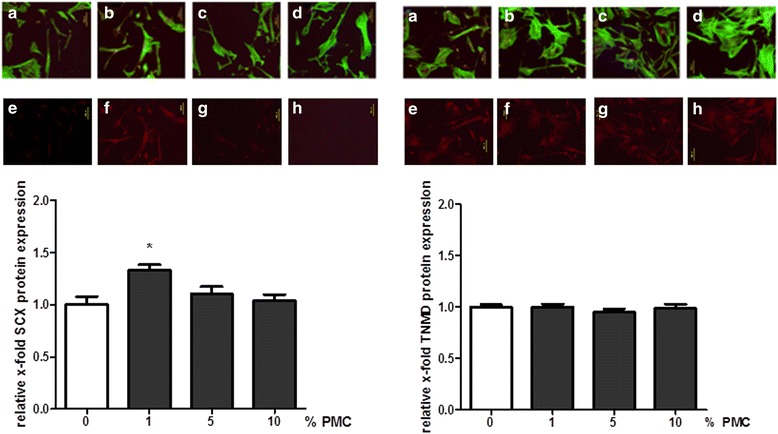
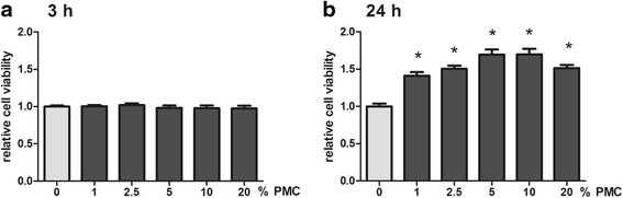
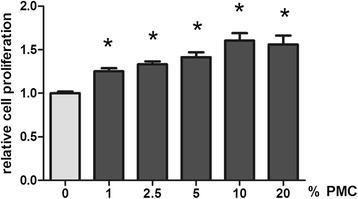
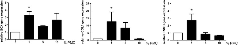
Using enzyme linked immunosorbent assay (ELISA) we were able to quantify the several growth factors and cytokines found in PMC. Tenocytes were isolated both from human and from mouse Achilles tendons and stimulated with PMC. CyQuant® and Cell Titer Blue® assays were carried out to analyze tendon growth and viability at different concentrations of PMC. Real time RT-PCR was used to analyze tenocyte gene expression with or without PMC treatment. Immunohistochemistry was carried out to detect the tenocyte-specific antibody tenomodulin (TNMD) and scleraxis (SCX).
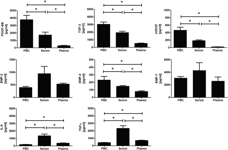
We were able to detect numerous mediators such as platelet derived growth factor BB (PDGF-BB), interleukin 6 (IL-6), vascular endothelial growth factor (VEGF), tumor necrosis factor (TNF-α), transforming growth factor beta 1 (TGF-ß1), and bone morphogenetic proteins 2, 4 and 7 (BMP-4, BMP-2, BMP-7) in PMC. It was possible to show a positive effect of PMC on human tendon cell growth and viability in a dose-dependent manner. Furthermore, PMC treatment led to induction of gene expression of scleraxis (SCX), type I collagen A 1 (Col1A1) and TNMD by tenocytes.
We suggest that the use of autologous PMC may be a suitable addition to conventional tendon therapy that is capable of increasing and optimizing tendon healing and reducing the risk of recurrence.
尽管有许多研究探讨了导致急性和慢性肌腱损伤的病因和病理因素,但肌腱损伤的病理生理学仍未完全明确。虽然大多数损伤并不复杂,但由于肌腱组织血管分布较差,治疗过程漫长且效果不尽人意。血小板介质浓缩物(PMC)含有许多源自血小板的生长因子,可促进伤口愈合。在本研究中,我们调查了PMC对肌腱细胞增殖和分化的影响,以便为组织再生策略提供实验依据并开发新的治疗理念。
使用酶联免疫吸附测定(ELISA),我们能够定量检测PMC中发现的几种生长因子和细胞因子。从人和小鼠的跟腱中分离出肌腱细胞,并用PMC进行刺激。采用CyQuant®和Cell Titer Blue®测定法分析不同浓度PMC下的肌腱生长和活力。使用实时逆转录聚合酶链反应(RT-PCR)分析有无PMC处理时肌腱细胞的基因表达。进行免疫组织化学检测肌腱细胞特异性抗体腱调蛋白(TNMD)和硬骨素(SCX)。
我们能够在PMC中检测到多种介质,如血小板衍生生长因子BB(PDGF-BB)、白细胞介素6(IL-6)、血管内皮生长因子(VEGF)、肿瘤坏死因子(TNF-α)、转化生长因子β1(TGF-β1)以及骨形态发生蛋白2、4和7(BMP-4、BMP-2、BMP-7)。结果表明,PMC对人肌腱细胞生长和活力具有剂量依赖性的积极作用。此外,PMC处理导致肌腱细胞诱导硬骨素(SCX)、I型胶原蛋白A1(Col1A1)和TNMD的基因表达。
我们认为,使用自体PMC可能是传统肌腱治疗的合适补充,能够增强和优化肌腱愈合并降低复发风险。