Sarro Lidia, Senjem Matthew L, Lundt Emily S, Przybelski Scott A, Lesnick Timothy G, Graff-Radford Jonathan, Boeve Bradley F, Lowe Val J, Ferman Tanis J, Knopman David S, Comi Giancarlo, Filippi Massimo, Petersen Ronald C, Jack Clifford R, Kantarci Kejal
1 Department of Radiology, Mayo Clinic, Rochester, MN, USA 2 Neuroimaging Research Unit, Institute of Experimental Neurology, Division of Neuroscience, San Raffaele Scientific Institute, Vita-Salute San Raffaele University, Milan, Italy 3 Department of Neurology, Institute of Experimental Neurology, Division of Neuroscience, San Raffaele Scientific Institute, Vita-Salute San Raffaele University, Milan, Italy.
1 Department of Radiology, Mayo Clinic, Rochester, MN, USA 4 Department of Information Technology, Mayo Clinic, Rochester, MN, USA.
Brain. 2016 Oct;139(Pt 10):2740-2750. doi: 10.1093/brain/aww193. Epub 2016 Jul 24.
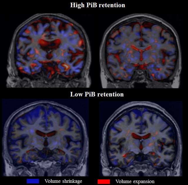
Alzheimer's disease pathology frequently coexists with Lewy body disease at autopsy in patients with probable dementia with Lewy bodies. More than half of patients with probable dementia with Lewy bodies have high amyloid-β deposition as measured with C-Pittsburgh compound B binding on positron emission tomography. Biomarkers of amyloid-β deposition precede neurodegeneration on magnetic resonance imaging during the progression of Alzheimer's disease, but little is known about how amyloid-β deposition relates to longitudinal progression of atrophy in patients with probable dementia with Lewy bodies. We investigated the associations between baseline C-Pittsburgh compound B binding on positron emission tomography and the longitudinal rates of grey matter atrophy in a cohort of clinically diagnosed patients with dementia with Lewy bodies (n = 20), who were consecutively recruited to the Mayo Clinic Alzheimer's Disease Research Centre. All patients underwent C-Pittsburgh compound B positron emission tomography and magnetic resonance imaging examinations at baseline. Follow-up magnetic resonance imaging was performed after a mean (standard deviation) interval of 2.5 (1.1) years. Regional grey matter loss was determined on three-dimensional T-weighted magnetic resonance imaging with the tensor-based morphometry-symmetric normalization technique. Linear regression was performed between baseline C-Pittsburgh compound B standard unit value ratio and longitudinal change in regional grey matter volumes from an in-house modified atlas. We identified significant associations between greater baseline C-Pittsburgh compound B standard unit value ratio and greater grey matter loss over time in the posterior cingulate gyrus, lateral and medial temporal lobe, and occipital lobe as well as caudate and putamen nuclei, after adjusting for age (P < 0.05). Greater baseline C-Pittsburgh compound B standard unit value ratio was also associated with greater ventricular expansion rates (P < 0.01) and greater worsening over time in Clinical Dementia Rating Scale, sum of boxes (P = 0.02). In conclusion, in patients with probable dementia with Lewy bodies, higher amyloid-β deposition at baseline is predictive of faster neurodegeneration in the cortex and also in the striatum. This distribution is suggestive of possible interactions among amyloid-β, tau and α-synuclein aggregates, which needs further investigation. Furthermore, higher amyloid-β deposition at baseline predicts a faster clinical decline over time in patients with probable dementia with Lewy bodies.
在可能患有路易体痴呆的患者尸检中,阿尔茨海默病病理常与路易体病共存。超过半数可能患有路易体痴呆的患者,经正电子发射断层扫描用C-匹兹堡化合物B结合法检测,显示有高淀粉样蛋白-β沉积。在阿尔茨海默病进展过程中,淀粉样蛋白-β沉积的生物标志物在磁共振成像上先于神经退行性变出现,但对于淀粉样蛋白-β沉积与可能患有路易体痴呆患者萎缩的纵向进展之间的关系,人们了解甚少。我们在梅奥诊所阿尔茨海默病研究中心连续招募的一组临床诊断为路易体痴呆的患者(n = 20)中,研究了基线正电子发射断层扫描时C-匹兹堡化合物B结合与灰质萎缩纵向速率之间的关联。所有患者在基线时均接受了C-匹兹堡化合物B正电子发射断层扫描和磁共振成像检查。平均(标准差)间隔2.5(1.1)年后进行了随访磁共振成像检查。采用基于张量的形态学对称归一化技术,在三维T加权磁共振成像上确定区域灰质损失。对基线C-匹兹堡化合物B标准单位价值比与根据内部改良图谱得出的区域灰质体积纵向变化进行线性回归分析。在调整年龄后,我们发现基线C-匹兹堡化合物B标准单位价值比越高,后扣带回、颞叶外侧和内侧以及枕叶以及尾状核和壳核随时间的灰质损失越大,差异有统计学意义(P < 0.05)。基线C-匹兹堡化合物B标准单位价值比越高,心室扩张率也越高(P < 0.01),且临床痴呆评定量表总分随时间恶化越严重(P = 0.02)。总之,在可能患有路易体痴呆的患者中,基线时较高的淀粉样蛋白-β沉积预示着皮质和纹状体中神经退行性变更快。这种分布提示淀粉样蛋白-β、tau和α-突触核蛋白聚集体之间可能存在相互作用,这需要进一步研究。此外,基线时较高的淀粉样蛋白-β沉积预示着可能患有路易体痴呆的患者随时间临床衰退更快。