State Key Laboratory of Military Stomatology, Department of Oral Anatomy and Physiology and TMD, School of Stomatology, Fourth Military Medical University, 145 Changle Western Road, Xi'an, 710032, China.
Department of Dentistry, Tangdu Hospital, Forth Military Medical University, Shannxi, Xi'an, 710038, China.
Sci Rep. 2016 Jul 25;6:30085. doi: 10.1038/srep30085.
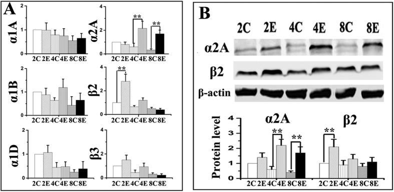
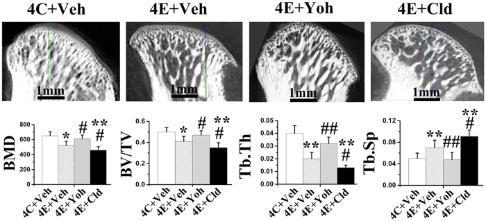
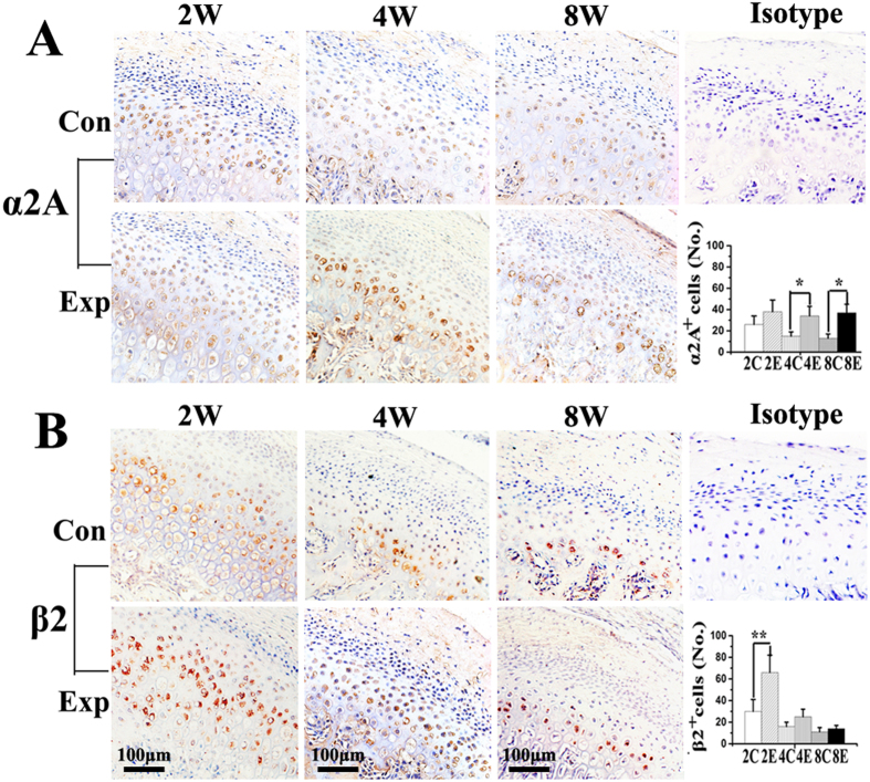
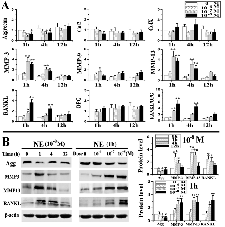
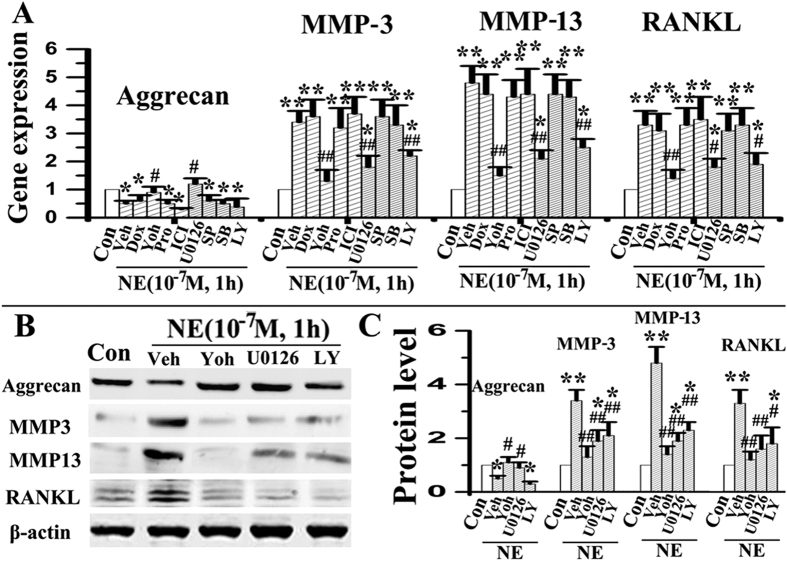
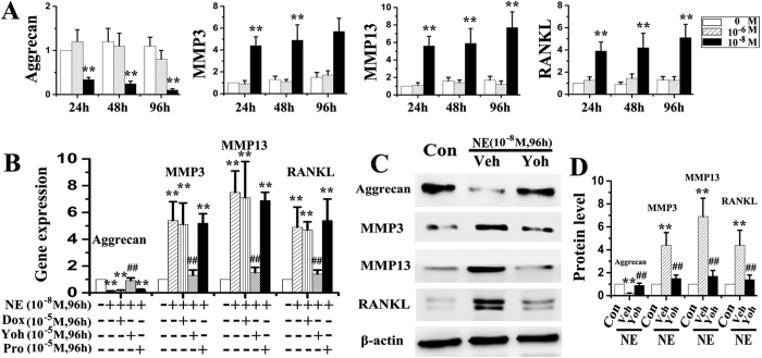
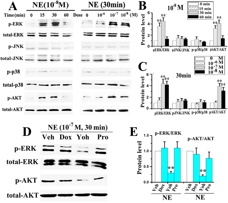
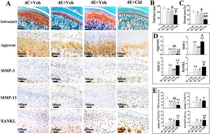
This study tested whether activation of adrenoreceptors in chondrocytes has roles in degenerative remodelling of temporomandibular joint (TMJ) and to determine associated mechanisms. Unilateral anterior crossbite (UAC) was established to induce TMJ degeneration in rats. Saline vehicle, α2- and β-adrenoreceptor antagonists or agonists were injected locally into the TMJ area of UAC rats. Cartilage degeneration, subchondral bone microarchitecture and the expression of adrenoreceptors, aggrecans, matrix metalloproteinases (MMPs) and RANKL by chondrocytes were evaluated. Chondrocytes were stimulated by norepinephrine to investigate signal transduction of adrenoreceptors. Increased α2A-adrenoreceptor expression was observed in condylar cartilage of UAC rats, together with cartilage degeneration and subchondral bone loss. Norepinephrine depresses aggrecans expression but stimulates MMP-3, MMP-13 and RANKL production by chondrocytes through ERK1/2 and PKA pathway; these effects were abolished by an α2A-adrenoreceptor antagonist. Furthermore, inhibition of α2A-adrenoreceptor attenuated degenerative remodelling in the condylar cartilage and subchondral bone, as revealed by increased cartilage thickness, proteoglycans and aggrecan expression, and decreased MMP-3, MMP-13 and RANKL expressions in cartilage, increased BMD, BV/TV, and decreased Tb.Sp in subchondral bone. Conversely, activation of α2A-adrenoreceptor intensified aforementioned degenerative changes in UAC rats. It is concluded that activation of α2A-adrenergic signal in chondrocytes promotes TMJ degenerative remodelling by chondrocyte-mediated pro-catabolic activities.
本研究旨在探讨软骨细胞肾上腺素能受体的激活在颞下颌关节(TMJ)退行性重塑中的作用及其相关机制。通过建立单侧前牙反合(UAC)大鼠模型来诱导 TMJ 退变。将生理盐水载体、α2-和β-肾上腺素能受体拮抗剂或激动剂局部注射到 UAC 大鼠的 TMJ 区域。评估软骨退变、软骨下骨微结构以及软骨细胞中肾上腺素能受体、聚集蛋白、基质金属蛋白酶(MMPs)和 RANKL 的表达。用去甲肾上腺素刺激软骨细胞,研究肾上腺素能受体的信号转导。结果发现,UAC 大鼠髁突软骨中α2A-肾上腺素能受体表达增加,同时伴有软骨退变和软骨下骨丢失。去甲肾上腺素通过 ERK1/2 和 PKA 通路抑制聚集蛋白的表达,但刺激 MMP-3、MMP-13 和 RANKL 的产生;这些作用被α2A-肾上腺素能受体拮抗剂所阻断。此外,抑制α2A-肾上腺素能受体可减轻髁突软骨和软骨下骨的退行性重塑,表现为软骨厚度、糖胺聚糖和聚集蛋白表达增加,软骨中 MMP-3、MMP-13 和 RANKL 表达减少,软骨下骨 BMD、BV/TV 增加,Tb.Sp 减少。相反,激活α2A-肾上腺素能受体则加剧了 UAC 大鼠的上述退行性变化。综上所述,软骨细胞中α2A-肾上腺素能信号的激活通过软骨细胞介导的促分解代谢活性促进 TMJ 退行性重塑。