State Key Laboratory of Military Stomatology & National Clinical Research Center for Oral Diseases & Shaanxi International Joint Research Center for Oral Diseases, Department of Oral Anatomy and Physiology and TMD, School of Stomatology, the Fourth Military Medical University, Xi'an, China.
Class 1, Grade 2018, School of Stomatology, Zhengzhou University, Zhengzhou, China.
Arthritis Res Ther. 2022 Feb 14;24(1):44. doi: 10.1186/s13075-022-02738-5.
Abnormal cartilage calcification is one of the pathological changes of temporomandibular joint (TMJ) osteoarthritis (OA). Recent studies have reported that exosomes can regulate the formation of abnormal calcified nodules in diseases including atherosclerosis and chronic kidney disease. However, the influences of chondrocyte-derived exosomes on abnormal cartilage calcification in TMJ OA are still unclear.
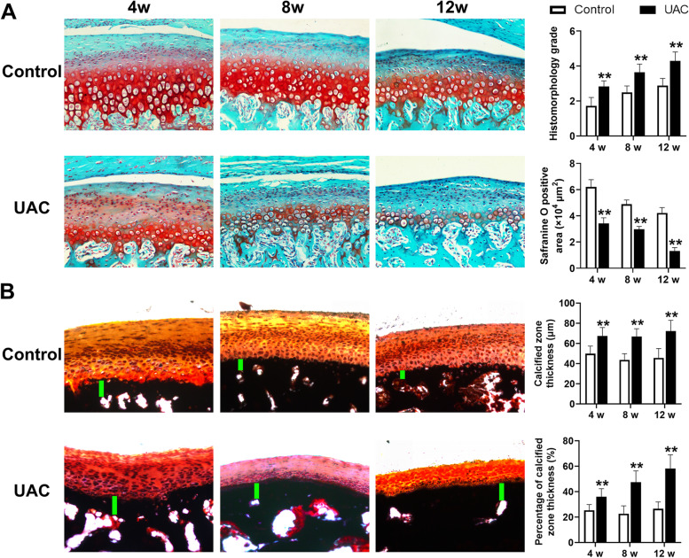
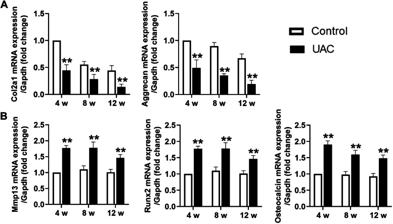
TMJ OA was induced by unilateral anterior crossbite (UAC) for 4, 8, or 12 weeks in rats to observe abnormal calcification in TMJ condylar cartilage and exosome formation. Concomitantly, GW4869, the inhibitor of exosome formation, was locally injected to the TMJ of rats under stimulation of UAC, while the exosomes extracted from primary condylar chondrocytes stimulated with fluid flow shear stress (FFSS) were locally injected to rats TMJ.
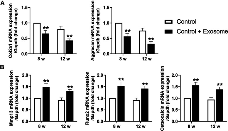
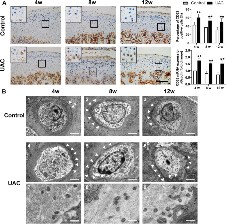
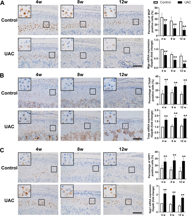
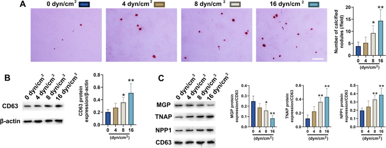
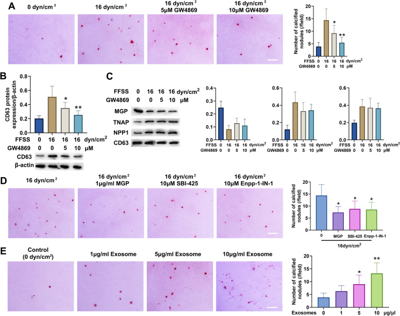
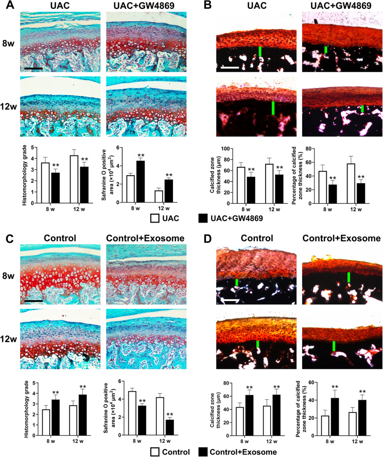
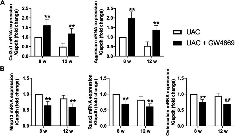
Abnormal calcification was enhanced in the degenerative cartilage of TMJ OA in UAC rats, and a large number of exosome-like structures with diameters of 50-150 nm were found in the calcified cartilage together with decreased expression of matrix Gla protein (MGP) and increased expression of CD63, tissue-nonspecific alkaline phosphatase (TNAP) and nucleotide pyrophosphatase/phosphodiesterase-1 (NPP1). After FFSS stimulation, the number of exosomes secreted by chondrocytes and the numbers of calcified nodules were increased in cultured cells, and the protein levels of MGP, TNAP, and NPP1 in exosomes were changed. Inhibition of exosome formation, TNAP, and NPP1 or supplementation with exogenous MGP effectively alleviated FFSS-induced chondrocyte calcification. Local injection of GW4869, the exosome inhibitor, alleviated TMJ OA-related cartilage degeneration and calcification in UAC rats. Local injection of exosomes obtained from chondrocytes stimulated by FFSS to the TMJs of normal rats induced cartilage degeneration and calcification similar to that in TMJ OA.
Abnormal biomechanical loading leads to enhanced formation of chondrocyte-derived exosomes, in which promoters of calcification increased and inhibitors decreased, resulting in accelerating abnormal cartilage calcification in TMJ OA. The inhibition of degenerative chondrocyte-derived exosomes is expected to be a new way to prevent and treat TMJ OA.
异常软骨钙化是颞下颌关节(TMJ)骨关节炎(OA)的病理变化之一。最近的研究报告称,外泌体可调节动脉粥样硬化和慢性肾脏病等疾病中异常钙化结节的形成。然而,软骨细胞来源的外泌体对 TMJ OA 中异常软骨钙化的影响尚不清楚。
通过单侧前牙反颌(UAC)诱导大鼠 TMJ OA 4、8 或 12 周,观察 TMJ 髁突软骨的异常钙化和外泌体的形成。同时,在 UAC 刺激下,将外泌体形成抑制剂 GW4869 局部注射到大鼠 TMJ 中,将流体剪切力(FFSS)刺激的原代髁状突软骨细胞提取的外泌体局部注射到大鼠 TMJ 中。
UAC 大鼠 TMJ OA 退变软骨中异常钙化增强,钙化软骨中发现大量直径为 50-150nm 的外泌体样结构,基质 Gla 蛋白(MGP)表达降低,CD63、组织非特异性碱性磷酸酶(TNAP)和核苷酸焦磷酸酶/磷酸二酯酶-1(NPP1)表达增加。FFSS 刺激后,培养细胞中软骨细胞分泌的外泌体数量和钙化结节数量增加,外泌体中 MGP、TNAP 和 NPP1 的蛋白水平发生变化。抑制外泌体形成、TNAP 和 NPP1 或补充外源性 MGP 可有效缓解 FFSS 诱导的软骨细胞钙化。外泌体抑制剂 GW4869 的局部注射可减轻 UAC 大鼠 TMJ OA 相关的软骨退变和钙化。FFSS 刺激的软骨细胞提取的外泌体局部注射到正常大鼠 TMJ 中,可诱导类似 TMJ OA 的软骨退变和钙化。
异常生物力学负荷导致软骨细胞来源的外泌体形成增加,其中促进钙化的物质增加,抑制钙化的物质减少,导致 TMJ OA 中异常软骨钙化加速。抑制退变软骨细胞来源的外泌体有望成为预防和治疗 TMJ OA 的新方法。