• 文献检索
  • 文档翻译
  • 深度研究
  • 学术资讯
  • Suppr Zotero 插件Zotero 插件
  • 邀请有礼
  • 套餐&价格
  • 历史记录
应用&插件
Suppr Zotero 插件Zotero 插件浏览器插件Mac 客户端Windows 客户端微信小程序
定价
高级版会员购买积分包购买API积分包
服务
文献检索文档翻译深度研究API 文档MCP 服务
关于我们
关于 Suppr公司介绍联系我们用户协议隐私条款
关注我们

Suppr 超能文献

核心技术专利:CN118964589B侵权必究
粤ICP备2023148730 号-1Suppr @ 2026

文献检索

告别复杂PubMed语法,用中文像聊天一样搜索,搜遍4000万医学文献。AI智能推荐,让科研检索更轻松。

立即免费搜索

文件翻译

保留排版,准确专业,支持PDF/Word/PPT等文件格式,支持 12+语言互译。

免费翻译文档

深度研究

AI帮你快速写综述,25分钟生成高质量综述,智能提取关键信息,辅助科研写作。

立即免费体验

正常受试者二维斑点追踪成像评估膈肌

Diaphragm assessment by two dimensional speckle tracking imaging in normal subjects.

作者信息

Orde Sam R, Boon Andrea J, Firth Daniel G, Villarraga Hector R, Sekiguchi Hiroshi

机构信息

Division of Cardiovascular Diseases, Mayo Clinic, Rochester, Minnesota, USA.

Department of Intensive Care, Nepean Hospital, Sydney, Australia.

出版信息

BMC Anesthesiol. 2016 Jul 25;16(1):43. doi: 10.1186/s12871-016-0201-6.

DOI:10.1186/s12871-016-0201-6
PMID:27456490
原文链接:https://pmc.ncbi.nlm.nih.gov/articles/PMC4960718/
Abstract

BACKGROUND

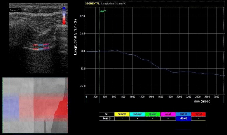
Conventionally, ultrasonographic assessment of diaphragm contractility has involved measuring respiratory changes in diaphragm thickness (thickening fraction) using B-mode or caudal displacement with M-mode. Two-dimensional speckle-tracking has been increasingly used to assess muscle deformation ('strain') in echocardiography. We sought to determine in a pilot study if this technology could be utilized to analyze diaphragmatic contraction.

METHODS

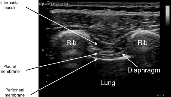
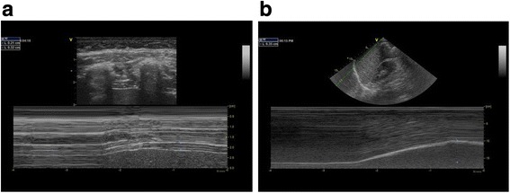
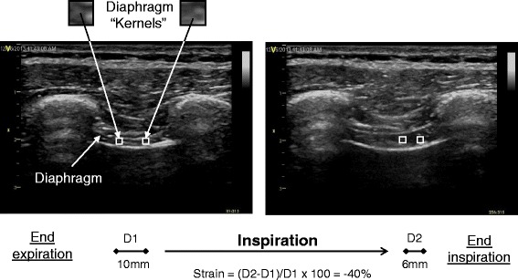
Fifty healthy adult volunteers with normal exercise capacity underwent ultrasound imaging. A linear array transducer was used for the assessment of diaphragm thickness, thickening fraction (TF), and strain in the right anterior axillary line at approximately the ninth intercostal space. A phased array transducer was applied subcostally for the assessment of diaphragm displacement on the right mid-clavicular line. Diaphragmatic images were recorded from the end of expiration through the end of inspiration at 60 % maximal inspiratory capacity. Diaphragm strain was analyzed off-line by speckle tracking imaging. Blinded inter- and intra-rater variability was tested in 10 cases.

RESULTS

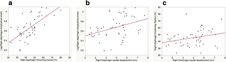
Mean right diaphragm thickness at end-expiration (±SD: standard deviation) was 0.24 cm (±0.1), with TF of 45.1 % (±12) at 60 % peak inspiratory effort. Mean right diaphragm caudal displacement was 4.9 cm (±1). Mean right diaphragm strain was -40.3 % (±9). A moderate correlation was seen between longitudinal strain and TF (R(2) 0.44, p < 0.0001). A weak correlation was seen between strain and caudal displacement (R(2) 0.14, p < 0.01), and an even weaker correlation was seen between caudal displacement and TF (R(2) 0.1, p = 0.04). Age, gender, and body mass index were not significantly associated with right diaphragm strain or TF. Although inter- and intra-rater variability was overall good for TF, caudal displacement, and strain (inter-rater R(2); 0.8, 0.9, and 0.7, respectively [p < 0.01], intra-rater R(2); 0.9, 0.7, and 0.9, respectively [p < 0.01]), strain values did have a slightly lower inter-rater repeatability.

CONCLUSIONS

Diaphragmatic strain estimated by speckle tracking imaging was associated with conventional ultrasound measures of diaphragmatic function (TF and caudal displacement). Further clinical studies are warranted to investigate its clinical utility.

摘要

背景

传统上,超声评估膈肌收缩功能涉及使用B模式测量膈肌厚度的呼吸变化(增厚率)或使用M模式测量膈肌尾端位移。二维斑点追踪技术在超声心动图中越来越多地用于评估心肌变形(“应变”)。我们试图在一项初步研究中确定该技术是否可用于分析膈肌收缩。

方法

50名运动能力正常的健康成年志愿者接受了超声成像检查。使用线性阵列换能器在右腋前线大约第9肋间评估膈肌厚度、增厚率(TF)和应变。使用相控阵换能器在右锁骨中线肋下评估膈肌位移。在60%最大吸气量时,从呼气末到吸气末记录膈肌图像。通过斑点追踪成像离线分析膈肌应变。对10例病例进行了评分者间和评分者内的盲法变异性测试。

结果

呼气末右膈肌平均厚度(±标准差)为0.24cm(±0.1),在60%吸气峰值时TF为45.1%(±12)。右膈肌平均尾端位移为4.9cm(±1)。右膈肌平均应变是-40.3%(±9)。纵向应变与TF之间存在中度相关性(R² 0.44,p<0.0001)。应变与尾端位移之间存在弱相关性(R² 0.14,p<0.01),尾端位移与TF之间的相关性更弱(R² 0.1,p = 0.04)。年龄、性别和体重指数与右膈肌应变或TF无显著相关性。虽然评分者间和评分者内变异性对于TF、尾端位移和应变总体良好(评分者间R²分别为0.8、0.9和0.7 [p<0.01],评分者内R²分别为0.9、0.7和0.9 [p<0.01]),但应变值的评分者间重复性略低。

结论

通过斑点追踪成像估计的膈肌应变与膈肌功能的传统超声测量指标(TF和尾端位移)相关。有必要进行进一步的临床研究以探讨其临床应用价值。

https://cdn.ncbi.nlm.nih.gov/pmc/blobs/a0d2/4960718/3d5e782296f6/12871_2016_201_Fig5_HTML.jpg
https://cdn.ncbi.nlm.nih.gov/pmc/blobs/a0d2/4960718/015123fc56a6/12871_2016_201_Fig1_HTML.jpg
https://cdn.ncbi.nlm.nih.gov/pmc/blobs/a0d2/4960718/8bce2aa91b66/12871_2016_201_Fig2_HTML.jpg
https://cdn.ncbi.nlm.nih.gov/pmc/blobs/a0d2/4960718/a55d639299d5/12871_2016_201_Fig3_HTML.jpg
https://cdn.ncbi.nlm.nih.gov/pmc/blobs/a0d2/4960718/7adab80ea4b3/12871_2016_201_Fig4_HTML.jpg
https://cdn.ncbi.nlm.nih.gov/pmc/blobs/a0d2/4960718/3d5e782296f6/12871_2016_201_Fig5_HTML.jpg

相似文献

1
Diaphragm assessment by two dimensional speckle tracking imaging in normal subjects.正常受试者二维斑点追踪成像评估膈肌
BMC Anesthesiol. 2016 Jul 25;16(1):43. doi: 10.1186/s12871-016-0201-6.
2
Functional assessment of the diaphragm by speckle tracking ultrasound during inspiratory loading.在吸气负荷下通过斑点追踪超声对膈肌进行功能评估。
J Appl Physiol (1985). 2017 Nov 1;123(5):1063-1070. doi: 10.1152/japplphysiol.00095.2017. Epub 2017 May 18.
3
Comparison of assessment of diaphragm function using speckle tracking between patients with successful and failed weaning: a multicentre, observational, pilot study.使用斑点追踪技术比较成功撤机与撤机失败患者的膈肌功能评估:一项多中心、观察性、先导研究。
BMC Pulm Med. 2022 Dec 1;22(1):459. doi: 10.1186/s12890-022-02260-z.
4
Diaphragmatic thickness-lung volume relationship in vivo.体内膈肌厚度与肺容积的关系。
J Appl Physiol (1985). 1989 Oct;67(4):1560-8. doi: 10.1152/jappl.1989.67.4.1560.
5
Reproducibility and Clinical Correlates of Supine Diaphragmatic Motion Measured by M-Mode Ultrasonography in Healthy Volunteers.仰卧位超声 M 模式测量健康志愿者膈肌运动的可重复性及其临床相关性。
Respiration. 2018;96(3):259-266. doi: 10.1159/000489229. Epub 2018 Aug 16.
6
Use of Angle-Independent M-Mode Sonography for Assessment of Diaphragm Displacement.使用角度无关型M型超声检查评估膈肌位移
J Ultrasound Med. 2016 Dec;35(12):2615-2621. doi: 10.7863/ultra.15.11100. Epub 2016 Oct 25.
7
Evaluation of diaphragm thickening by diaphragm ultrasonography: a reproducibility and a repeatability study.膈肌超声评估膈肌增厚:一项可重复性和再现性研究。
J Ultrasound. 2021 Dec;24(4):411-416. doi: 10.1007/s40477-020-00462-x. Epub 2020 May 1.
8
Ultrasonographic measurement of diaphragm muscle activity for assessment of respiratory function in horses.超声测量膈肌肌电活动评估马的呼吸功能。
J Equine Vet Sci. 2024 Jul;138:105100. doi: 10.1016/j.jevs.2024.105100. Epub 2024 May 18.
9
Reference values of diaphragmatic dimensions in healthy children aged 0-8 years.健康 0-8 岁儿童膈肌尺寸参考值。
Eur J Pediatr. 2023 Jun;182(6):2577-2589. doi: 10.1007/s00431-023-04920-6. Epub 2023 Mar 20.
10
A novel application for assessing diaphragmatic function by ultrasonic deformation analysis in noninvasively ventilated healthy young adults.一种通过超声变形分析评估无创通气健康年轻成年人膈肌功能的新应用。
Ultraschall Med. 2014 Dec;35(6):540-6. doi: 10.1055/s-0034-1366090. Epub 2014 Mar 19.

引用本文的文献

1
Diaphragmatic ultrasound and its relationship to breathing effort and load: a prospective observational study.膈肌超声及其与呼吸功和负荷的关系:一项前瞻性观察性研究。
Crit Care. 2025 May 13;29(1):190. doi: 10.1186/s13054-025-05436-1.
2
Advancements in imaging techniques for monitoring the respiratory muscles.用于监测呼吸肌的成像技术进展。
Crit Care. 2025 Mar 12;29(1):110. doi: 10.1186/s13054-025-05339-1.
3
Speckle tracking ultrasound as a new tool to predict the weaning outcome of mechanical ventilation patients: a prospective observational study.

本文引用的文献

1
Diaphragm ultrasound as indicator of respiratory effort in critically ill patients undergoing assisted mechanical ventilation: a pilot clinical study.膈肌超声作为接受机械通气的危重症患者呼吸努力的指标:一项初步临床研究。
Crit Care. 2015 Apr 13;19(1):161. doi: 10.1186/s13054-015-0894-9.
2
Intensive care ultrasound: III. Lung and pleural ultrasound for the intensivist.重症监护超声:III. 重症监护医生的肺部和胸膜超声检查
Ann Am Thorac Soc. 2013 Dec;10(6):708-12. doi: 10.1513/AnnalsATS.201308-288OT.
3
Diaphragm breathing movement measurement using ultrasound and radiographic imaging: a concurrent validity.
斑点追踪超声作为预测机械通气患者撤机结局的新工具:一项前瞻性观察性研究。
Front Med (Lausanne). 2024 Dec 6;11:1449938. doi: 10.3389/fmed.2024.1449938. eCollection 2024.
4
Diaphragm ultrasound and diaphragmatic 2D speckle tracking imaging in acute heart failure: a case series.急性心力衰竭中的膈肌超声与膈肌二维斑点追踪成像:病例系列
Eur Heart J Case Rep. 2024 Nov 28;8(12):ytae632. doi: 10.1093/ehjcr/ytae632. eCollection 2024 Dec.
5
Decreased diaphragm moving distance measured by ultrasound speckle tracking reflects poor prognosis in amyotrophic lateral sclerosis.通过超声斑点追踪测量的膈肌移动距离减小反映了肌萎缩侧索硬化症的预后不良。
Clin Neurophysiol Pract. 2024 Oct 22;9:252-260. doi: 10.1016/j.cnp.2024.10.002. eCollection 2024.
6
Comparative Effect of High-Frequency Nasal Cannula and Noninvasive Ventilation on the Work of Breathing and Postoperative Pulmonary Complication after Pediatric Congenital Cardiac Surgery: A Prospective Randomized Controlled Trial.高频鼻导管与无创通气对小儿先天性心脏病术后呼吸做功及肺部并发症的影响:一项前瞻性随机对照试验。
Ann Card Anaesth. 2024 Jan 1;27(1):43-50. doi: 10.4103/aca.aca_130_23. Epub 2024 Jan 12.
7
Perioperative Diaphragm Dysfunction.围手术期膈肌功能障碍
J Clin Med. 2024 Jan 17;13(2):519. doi: 10.3390/jcm13020519.
8
Replacement of fluoroscopy by ultrasonography in the evaluation of hemidiaphragm function, an exploratory prospective study.超声检查替代荧光透视用于评估半膈肌功能的探索性前瞻性研究。
Ultrasound J. 2024 Jan 8;16(1):1. doi: 10.1186/s13089-023-00355-0.
9
Reliability and validity of ultrasonography in evaluating the thickness, excursion, stiffness, and strain rate of respiratory muscles in non-hospitalized individuals: a systematic review.超声评估非住院个体呼吸肌厚度、活动度、僵硬度和应变率的可靠性和有效性:系统评价。
BMC Oral Health. 2023 Dec 2;23(1):959. doi: 10.1186/s12903-023-03558-y.
10
Ultrasound assessment of diaphragm thickness, contractility, and strain in healthy pediatric patients.超声评估健康儿科患者的膈肌厚度、收缩性和应变。
Pediatr Pulmonol. 2024 Feb;59(2):433-441. doi: 10.1002/ppul.26768. Epub 2023 Dec 1.
使用超声和放射成像测量膈肌呼吸运动:一种同时效度。
Biomed Mater Eng. 2014;24(1):947-52. doi: 10.3233/BME-130889.
4
Variability in diaphragm motion during normal breathing, assessed with B-mode ultrasound.正常呼吸时膈肌运动的变化,采用 B 型超声评估。
J Orthop Sports Phys Ther. 2013 Dec;43(12):927-31. doi: 10.2519/jospt.2013.4931. Epub 2013 Oct 11.
5
Two-dimensional strain ultrasound speckle tracking as a novel approach for the evaluation of right hemidiaphragmatic longitudinal deformation.二维应变超声斑点追踪技术作为评估右半膈肌纵向变形的一种新方法。
Exp Ther Med. 2013 Aug;6(2):368-372. doi: 10.3892/etm.2013.1133. Epub 2013 May 30.
6
Normality tests for statistical analysis: a guide for non-statisticians.用于统计分析的正态性检验:非统计学家指南。
Int J Endocrinol Metab. 2012 Spring;10(2):486-9. doi: 10.5812/ijem.3505. Epub 2012 Apr 20.
7
Two-dimensional ultrasound imaging of the diaphragm: quantitative values in normal subjects.二维超声膈肌成像:正常受试者的定量值。
Muscle Nerve. 2013 Jun;47(6):884-9. doi: 10.1002/mus.23702. Epub 2013 Apr 29.
8
From speckle tracking echocardiography to torsion: research tool today, clinical practice tomorrow.从斑点追踪超声心动图到扭转:今天的研究工具,明天的临床实践。
Curr Opin Crit Care. 2013 Jun;19(3):250-7. doi: 10.1097/MCC.0b013e32836092b7.
9
Ultrasound in critically ill patients: focus on diaphragm.
Intensive Care Med. 2013 May;39(5):986. doi: 10.1007/s00134-013-2882-3. Epub 2013 Mar 16.
10
Sonographic evaluation of the diaphragm in critically ill patients. Technique and clinical applications.超声评估危重症患者的膈肌:技术与临床应用。
Intensive Care Med. 2013 May;39(5):801-10. doi: 10.1007/s00134-013-2823-1. Epub 2013 Jan 24.