Pastorek Lukáš, Sobol Margarita, Hozák Pavel
Department of Biology of the Cell Nucleus, Institute of Molecular Genetics ASCR v.v.i., Vídeňská 1083, 142 20, Prague 4, Czech Republic.
Microscopy Centre, Institute of Molecular Genetics ASCR v.v.i., Vídeňská 1083, 142 20, Prague 4, Czech Republic.
Histochem Cell Biol. 2016 Oct;146(4):391-406. doi: 10.1007/s00418-016-1467-y. Epub 2016 Jul 26.
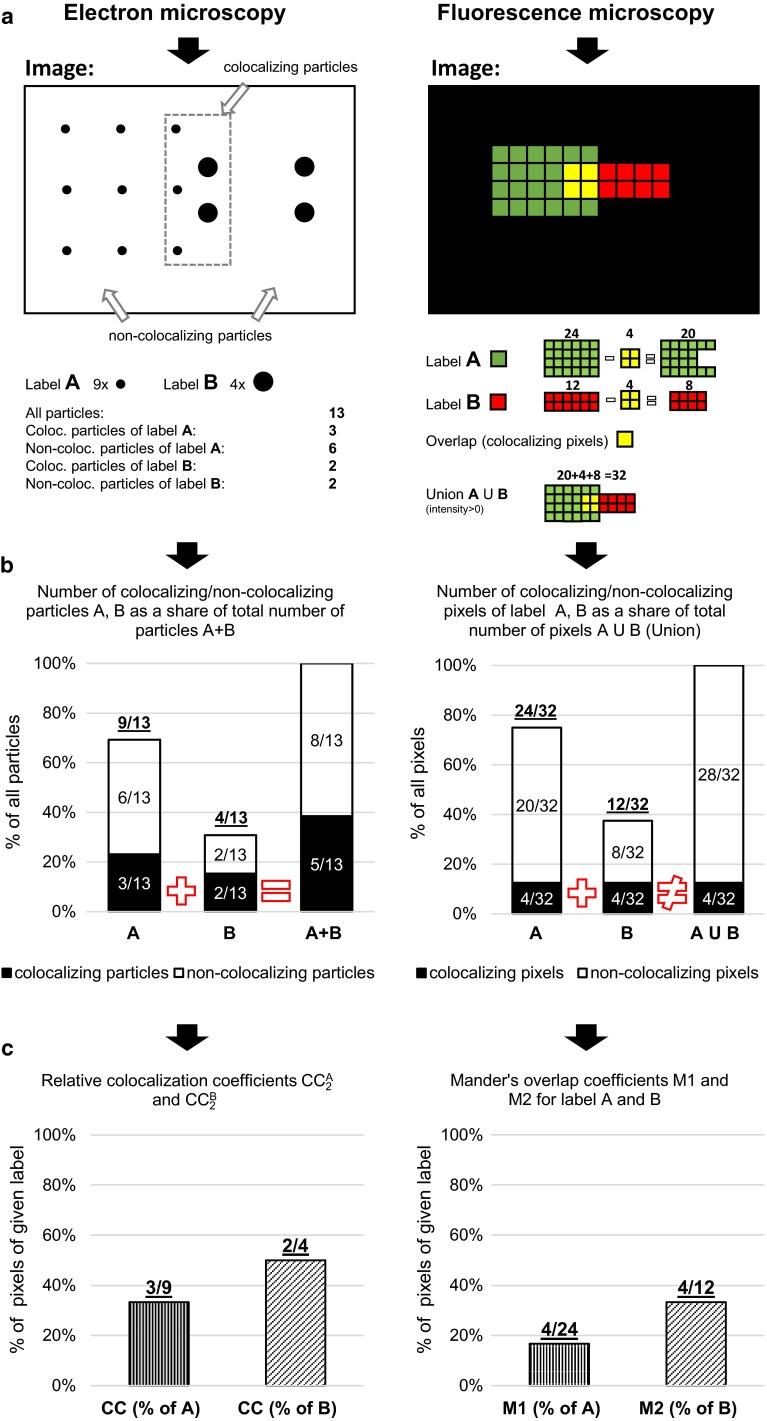
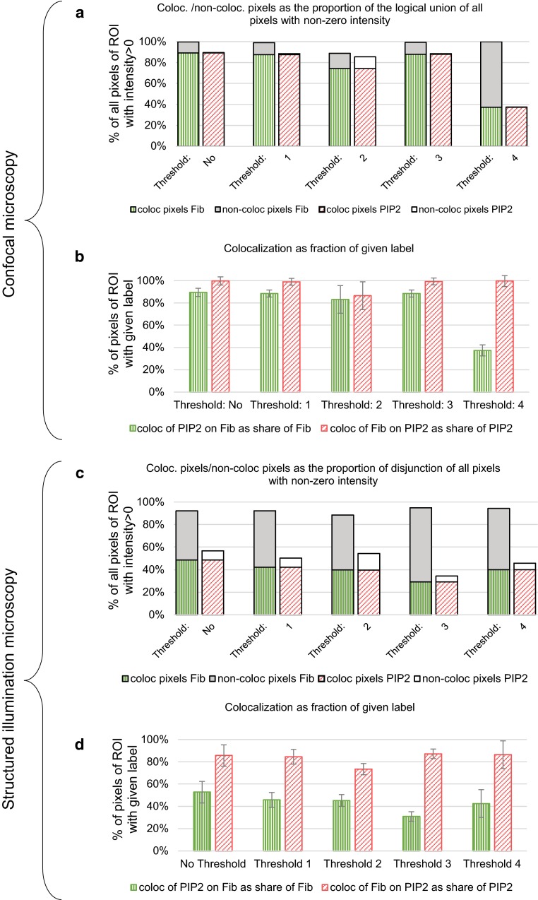
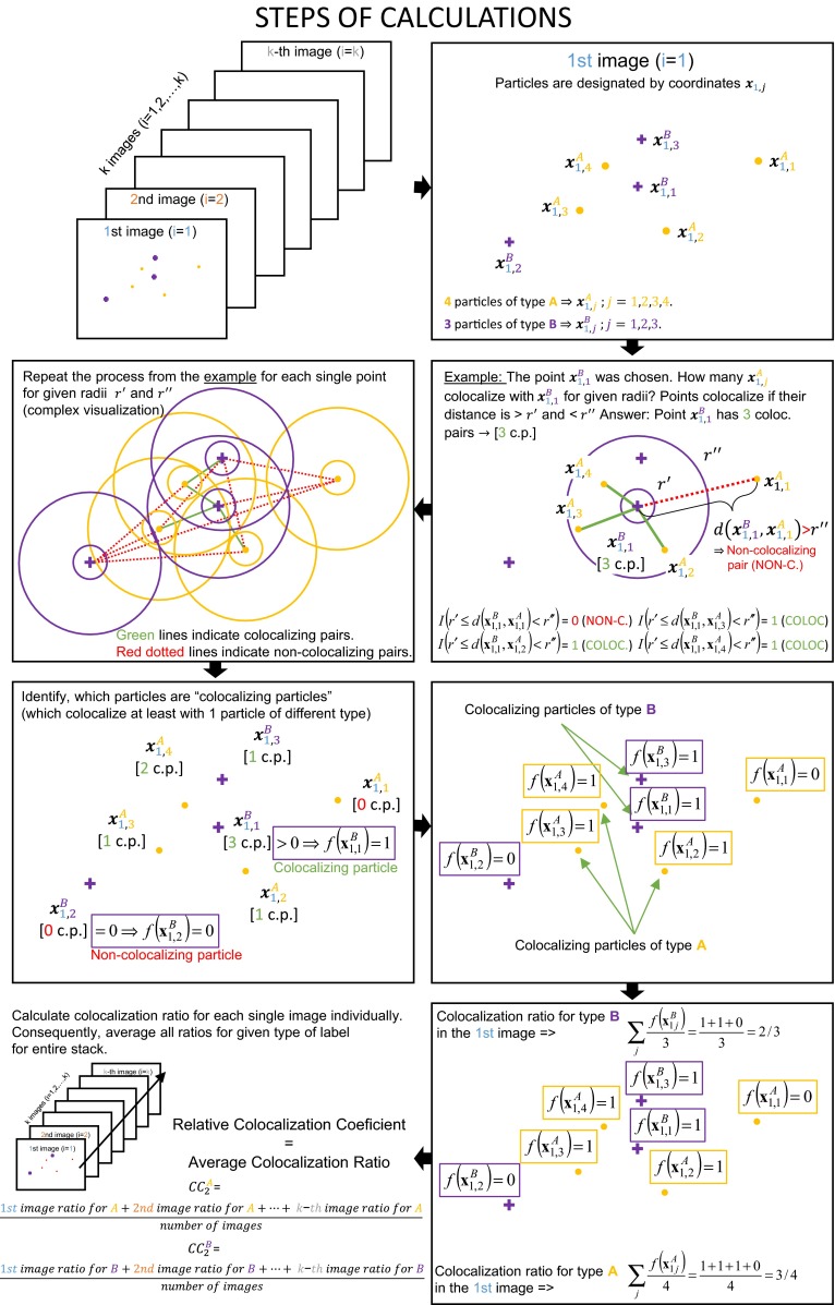
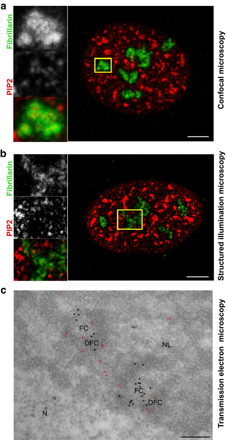
In biomedical studies, the colocalization is commonly understood as the overlap between distinctive labelings in images. This term is usually associated especially with quantitative evaluation of the immunostaining in fluorescence microscopy. On the other hand, the evaluation of the immunolabeling colocalization in the electron microscopy images is still under-investigated and biased by the subjective and non-quantitative interpretation of the image data. We introduce a novel computational technique for quantifying the level of colocalization in pointed patterns. Our approach follows the idea included in the widely used Manders' colocalization coefficients in fluorescence microscopy and represents its counterpart for electron microscopy. In presented methodology, colocalization is understood as the product of the spatial interactions at the single-particle (single-molecule) level. Our approach extends the current significance testing in the immunoelectron microscopy images and establishes the descriptive colocalization coefficients. To demonstrate the performance of the proposed coefficients, we investigated the level of spatial interactions of phosphatidylinositol 4,5-bisphosphate with fibrillarin in nucleoli. We compared the electron microscopy colocalization coefficients with Manders' colocalization coefficients for confocal microscopy and super-resolution structured illumination microscopy. The similar tendency of the values obtained using different colocalization approaches suggests the biological validity of the scientific conclusions. The presented methodology represents a good basis for further development of the quantitative analysis of immunoelectron microscopy data and can be used for studying molecular interactions at the ultrastructural level. Moreover, this methodology can be applied also to the other super-resolution microscopy techniques focused on characterization of discrete pointed structures.
在生物医学研究中,共定位通常被理解为图像中不同标记之间的重叠。这个术语通常特别与荧光显微镜中免疫染色的定量评估相关。另一方面,电子显微镜图像中免疫标记共定位的评估仍未得到充分研究,并且受到图像数据主观和非定量解释的影响。我们引入了一种新颖的计算技术来量化点状模式中的共定位水平。我们的方法遵循了荧光显微镜中广泛使用的曼德尔斯共定位系数所包含的思想,并代表了其在电子显微镜中的对应方法。在提出的方法中,共定位被理解为单粒子(单分子)水平上空间相互作用的产物。我们的方法扩展了当前免疫电子显微镜图像中的显著性测试,并建立了描述性共定位系数。为了证明所提出系数的性能,我们研究了磷脂酰肌醇4,5-二磷酸与核仁中纤维蛋白原的空间相互作用水平。我们将电子显微镜共定位系数与共聚焦显微镜和超分辨率结构光照显微镜的曼德尔斯共定位系数进行了比较。使用不同共定位方法获得的值的相似趋势表明了科学结论的生物学有效性。所提出的方法为免疫电子显微镜数据定量分析的进一步发展奠定了良好基础,可用于研究超微结构水平上的分子相互作用。此外,该方法也可应用于其他专注于离散点状结构表征的超分辨率显微镜技术。