Ruddy Kathy L, Leemans Alexander, Carson Richard G
School of Psychology, Queen's University Belfast, Belfast, BT7 1NN, UK.
Trinity College Institute of Neuroscience and School of Psychology, Trinity College Dublin, Dublin, Ireland.
Brain Struct Funct. 2017 Apr;222(3):1243-1252. doi: 10.1007/s00429-016-1274-1. Epub 2016 Jul 28.
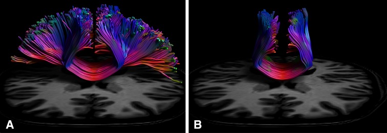
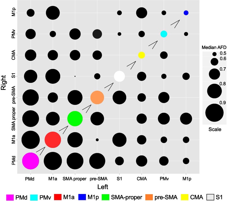
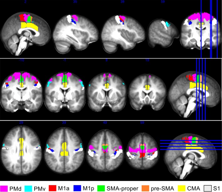
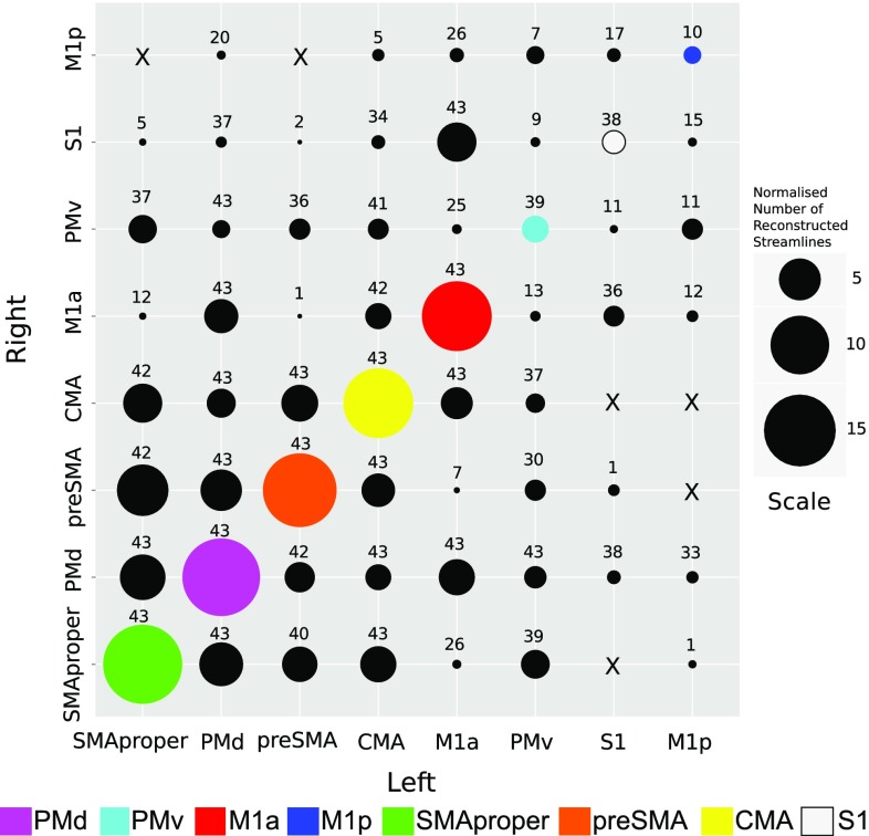
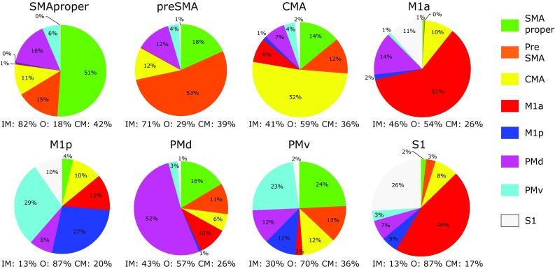
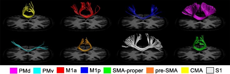
The organisational and architectural configuration of white matter pathways connecting brain regions has ramifications for all facets of the human condition, including manifestations of incipient neurodegeneration. Although diffusion tensor imaging (DTI) has been used extensively to visualise white matter connectivity, due to the widespread presence of crossing fibres, the lateral projections of the corpus callosum are not normally detected using this methodology. Detailed knowledge of the transcallosal connectivity of the human cortical motor network has, therefore, remained elusive. We employed constrained spherical deconvolution (CSD) tractography-an approach that is much less susceptible to the influence of crossing fibres, in order to derive complete in vivo characterizations of white matter pathways connecting specific motor cortical regions to their counterparts and other loci in the opposite hemisphere. The revealed patterns of connectivity closely resemble those derived from anatomical tracing in primates. It was established that dorsal premotor cortex (PMd) and supplementary motor area (SMA) have extensive interhemispheric connectivity-exhibiting both dense homologous projections, and widespread structural relations with every other region in the contralateral motor network. Through this in vivo portrayal, the importance of non-primary motor regions for interhemispheric communication is emphasised. Additionally, distinct connectivity profiles were detected for the anterior and posterior subdivisions of primary motor cortex. The present findings provide a comprehensive representation of transcallosal white matter projections in humans, and have the potential to inform the development of models and hypotheses relating structural and functional brain connectivity.
连接脑区的白质通路的组织和结构配置对人类状况的各个方面都有影响,包括早期神经退行性变的表现。尽管扩散张量成像(DTI)已被广泛用于可视化白质连接,但由于交叉纤维的广泛存在,通常无法使用这种方法检测胼胝体的外侧投射。因此,人类皮质运动网络的胼胝体连接的详细知识仍然难以捉摸。我们采用了约束球面去卷积(CSD)纤维束成像——一种对交叉纤维影响不太敏感的方法,以便在体内完整地表征连接特定运动皮质区域与其对侧半球对应区域及其他位点的白质通路。所揭示的连接模式与从灵长类动物解剖追踪中得出的模式非常相似。已确定背侧运动前区(PMd)和辅助运动区(SMA)具有广泛的半球间连接——既表现出密集的同源投射,又与对侧运动网络中的其他每个区域存在广泛的结构关系。通过这种体内描绘,强调了非初级运动区域在半球间通信中的重要性。此外,还检测到了初级运动皮质前后亚区的不同连接模式。本研究结果全面展示了人类胼胝体白质投射情况,并有潜力为有关脑结构和功能连接的模型及假设的发展提供信息。