Anzellotti Francesca, Onofrj Marco, Bonanni Laura, Saracino Antonio, Franciotti Raffaella
Department of Neuroscience, Imaging and Clinical Sciences, "G. d'Annunzio" University and Aging Research Centre, Ce.S.I., "G. d'Annunzio" University Foundation, Chieti, Italy.
Department of Neuroscience, Imaging and Clinical Sciences, "G. d'Annunzio" University and Aging Research Centre, Ce.S.I., "G. d'Annunzio" University Foundation, Chieti, Italy; Department of Neurology, SS Annunziata Hospital, Chieti, Italy.
Neuroimage Clin. 2016 Jul 2;12:212-8. doi: 10.1016/j.nicl.2016.07.001. eCollection 2016.
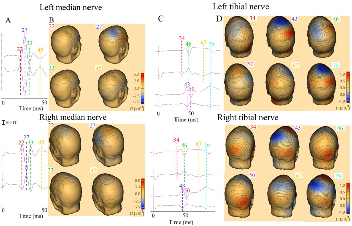
Enlarged cortical components of somatosensory evoked potentials (giant SEPs) recorded by electroencephalography (EEG) and abnormal somatosensory evoked magnetic fields (SEFs) recorded by magnetoencephalography (MEG) are observed in the majority of patients with cortical myoclonus (CM). Studies on simultaneous recordings of SEPs and SEFs showed that generator mechanism of giant SEPs involves both primary sensory and motor cortices. However the generator sources of giant SEPs have not been fully understood as only one report describes clearly giant SEPs following lower limb stimulation. In our study we performed a combined EEG-MEG recording on responses elicited by electric median and tibial nerve stimulation in a patient who developed consequently to methyl bromide intoxication CM with giant SEPs to median and tibial nerve stimuli. SEPs wave shapes were identified on the basis of polarity-latency components (e.g. P15-N20-P25) as defined by earlier studies and guidelines. At EEG recording, the SEP giant component did not appear in the latency range of the first cortical component for median nerve SEP (N20), but appeared instead in the range of the P37 tibial nerve SEP, which is currently identified as the first cortical component elicited by tibial nerve stimuli. Our MEG and EEG SEPs recordings also showed that components in the latency range of P37 were preceded by other cortical components. These findings suggest that lower limb P37 does not correspond to upper limb N20. MEG results confirmed that giant SEFs are the second component from both tibial (N43m-P43m) and median (N27m-P27m) nerve stimulation. MEG dipolar sources of these giant components were located in the primary sensory and motor area.
在大多数皮质肌阵挛(CM)患者中,可观察到通过脑电图(EEG)记录的体感诱发电位的皮质成分增大(巨大SEP),以及通过脑磁图(MEG)记录的异常体感诱发磁场(SEF)。对SEP和SEF同步记录的研究表明,巨大SEP的产生机制涉及初级感觉皮层和运动皮层。然而,巨大SEP的产生源尚未完全明确,因为仅有一篇报告清晰描述了下肢刺激后的巨大SEP。在我们的研究中,我们对一名因甲基溴中毒而发生CM且对正中神经和胫神经刺激出现巨大SEP的患者,进行了EEG - MEG联合记录,记录由正中神经和胫神经电刺激所引发的反应。SEP波形是根据早期研究和指南所定义的极性 - 潜伏期成分(如P15 - N20 - P25)来识别的。在EEG记录中,正中神经SEP的巨大成分未出现在第一个皮质成分(N20)的潜伏期范围内,而是出现在胫神经SEP的P37范围内,目前P37被确定为胫神经刺激所引发的第一个皮质成分。我们的MEG和EEG SEP记录还显示,P37潜伏期范围内的成分之前还有其他皮质成分。这些发现表明,下肢的P37与上肢的N20并不对应。MEG结果证实,巨大SEF是胫神经(N43m - P43m)和正中神经(N27m - P27m)刺激后的第二个成分。这些巨大成分的MEG偶极源位于初级感觉区和运动区。