• 文献检索
  • 文档翻译
  • 深度研究
  • 学术资讯
  • Suppr Zotero 插件Zotero 插件
  • 邀请有礼
  • 套餐&价格
  • 历史记录
应用&插件
Suppr Zotero 插件Zotero 插件浏览器插件Mac 客户端Windows 客户端微信小程序
定价
高级版会员购买积分包购买API积分包
服务
文献检索文档翻译深度研究API 文档MCP 服务
关于我们
关于 Suppr公司介绍联系我们用户协议隐私条款
关注我们

Suppr 超能文献

核心技术专利:CN118964589B侵权必究
粤ICP备2023148730 号-1Suppr @ 2026

文献检索

告别复杂PubMed语法,用中文像聊天一样搜索,搜遍4000万医学文献。AI智能推荐,让科研检索更轻松。

立即免费搜索

文件翻译

保留排版,准确专业,支持PDF/Word/PPT等文件格式,支持 12+语言互译。

免费翻译文档

深度研究

AI帮你快速写综述,25分钟生成高质量综述,智能提取关键信息,辅助科研写作。

立即免费体验

支气管内超声弹性成像。

Endobronchial ultrasound elastography.

机构信息

Department of Internal Medicine 2, Caritas Krankenhaus Bad Mergentheim, Uhlandstr, 7, 97980 Bad Mergentheim, Germany.

Department of Internal Medicine, Krankenhaus Märkisch Oderland, 15344 Strausberg, Germany.

出版信息

Endosc Ultrasound. 2016 Jul-Aug;5(4):233-8. doi: 10.4103/2303-9027.187866.

DOI:10.4103/2303-9027.187866
PMID:27503154
原文链接:https://pmc.ncbi.nlm.nih.gov/articles/PMC4989403/
Abstract

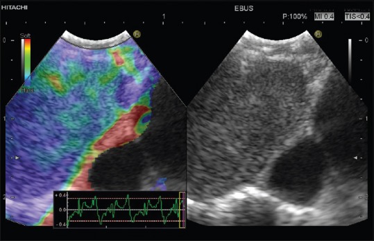
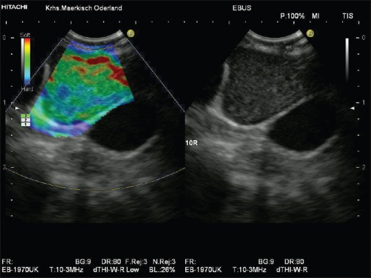
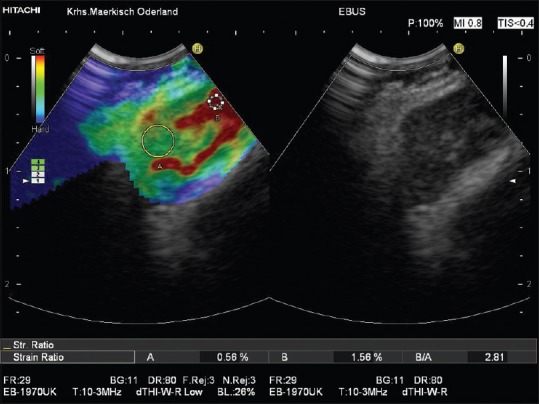
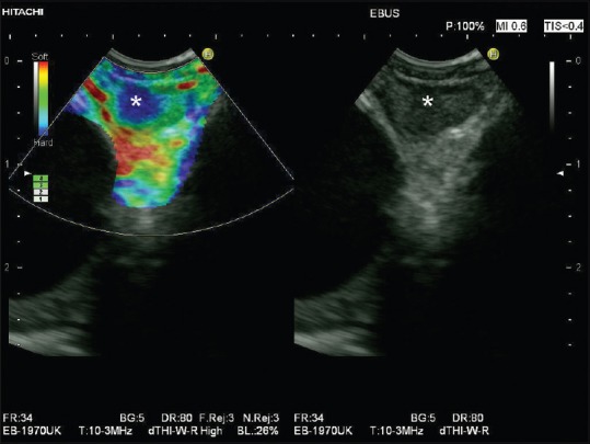
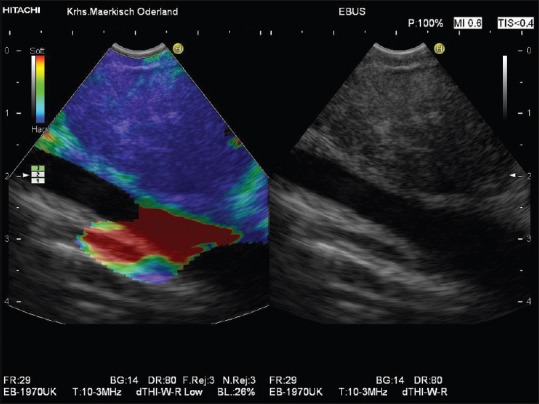
Elastographic techniques have recently become available as advanced diagnostic tools for tissue characterization. Strain elastography is a real-time technique used with transcutaneous ultrasound (US) and endoscopic US. Convincing evidence is available demonstrating a significant value of strain elastography for the discrimination of benign and malignant lymph nodes (LNs). This paper reviews preliminary data demonstrating the feasibility of performing real-time elastography during endobronchial US (EBUS) and a potential application of this technique for selection of LNs for EBUS-guided transbronchial needle aspiration in patients with lung cancer and extrathoracic malignancies.

摘要

弹性成像技术最近已成为用于组织特征描述的先进诊断工具。应变成像技术是一种实时技术,与经皮超声(US)和内镜超声(US)一起使用。有令人信服的证据表明,应变成像技术在鉴别良性和恶性淋巴结(LNs)方面具有重要价值。本文综述了初步数据,证明了在支气管内超声(EBUS)期间进行实时弹性成像的可行性,以及该技术在选择肺癌和胸外恶性肿瘤患者的 EBUS 引导经支气管针吸活检的 LN 方面的潜在应用。

https://cdn.ncbi.nlm.nih.gov/pmc/blobs/e0c5/4989403/184f2db9e5b8/EUS-5-233-g006.jpg
https://cdn.ncbi.nlm.nih.gov/pmc/blobs/e0c5/4989403/f69cbd9200f4/EUS-5-233-g001.jpg
https://cdn.ncbi.nlm.nih.gov/pmc/blobs/e0c5/4989403/0bce68856110/EUS-5-233-g002.jpg
https://cdn.ncbi.nlm.nih.gov/pmc/blobs/e0c5/4989403/c387cb35ed0b/EUS-5-233-g003.jpg
https://cdn.ncbi.nlm.nih.gov/pmc/blobs/e0c5/4989403/ff8809947c55/EUS-5-233-g004.jpg
https://cdn.ncbi.nlm.nih.gov/pmc/blobs/e0c5/4989403/f6607c37a1fd/EUS-5-233-g005.jpg
https://cdn.ncbi.nlm.nih.gov/pmc/blobs/e0c5/4989403/184f2db9e5b8/EUS-5-233-g006.jpg

相似文献

1
Endobronchial ultrasound elastography.支气管内超声弹性成像。
Endosc Ultrasound. 2016 Jul-Aug;5(4):233-8. doi: 10.4103/2303-9027.187866.
2
Diagnostic value of endobronchial ultrasound elastography for the differentiation of benign and malignant intrathoracic lymph nodes.经支气管超声弹性成像对鉴别良恶性胸内淋巴结的诊断价值。
Respirology. 2017 Jul;22(5):972-977. doi: 10.1111/resp.12979. Epub 2017 Jan 19.
3
Differentiating malignant and benign lymph nodes using endobronchial ultrasound elastography.使用支气管内超声弹性成像鉴别良恶性淋巴结。
J Formos Med Assoc. 2019 Jan;118(1 Pt 3):436-443. doi: 10.1016/j.jfma.2018.06.021. Epub 2018 Jul 12.
4
The combination of endobronchial elastography and sonographic findings during endobronchial ultrasound-guided transbronchial needle aspiration for predicting nodal metastasis.经支气管超声引导经支气管针吸活检术时弹性成像与超声表现联合预测淋巴结转移。
Thorac Cancer. 2019 Oct;10(10):2000-2005. doi: 10.1111/1759-7714.13186. Epub 2019 Sep 1.
5
Effectiveness of the Benign and Malignant Diagnosis of Mediastinal and Hilar Lymph Nodes by Endobronchial Ultrasound Elastography.经支气管超声弹性成像对纵隔及肺门淋巴结良恶性诊断的效能
J Cancer. 2017 Jul 2;8(10):1843-1848. doi: 10.7150/jca.19819. eCollection 2017.
6
Endobronchial ultrasound elastography in the diagnosis of mediastinal and hilar lymph nodes.经支气管超声弹性成像在纵隔和肺门淋巴结诊断中的应用。
Jpn J Clin Oncol. 2014 Oct;44(10):956-62. doi: 10.1093/jjco/hyu105. Epub 2014 Aug 13.
7
Endobronchial Ultrasound Elastography for Diagnosing Mediastinal and Hilar Lymph Nodes.支气管内超声弹性成像在诊断纵隔及肺门淋巴结中的应用
Chin Med J (Engl). 2015 Oct 20;128(20):2720-5. doi: 10.4103/0366-6999.167296.
8
Endobronchial Ultrasound Elastography for Evaluation of Intrathoracic Lymph Nodes: A Pilot Study.经支气管超声弹性成像评估胸内淋巴结:一项初步研究。
Respiration. 2017;93(5):327-338. doi: 10.1159/000464253. Epub 2017 Mar 22.
9
[Value of endobronchial ultrasound elastography in the diagnosis of mediastinal and hilar lymph node metastasis in lung cancer].[支气管内超声弹性成像在肺癌纵隔及肺门淋巴结转移诊断中的价值]
Zhong Nan Da Xue Xue Bao Yi Xue Ban. 2016 Jan;41(1):30-6. doi: 10.11817/j.issn.1672-7347.2016.01.005.
10
Contribution of endobronchial ultrasound elastography to the characterization of mediastinal lymphadenopathy: A single-center, prospective, observational study.经支气管超声弹性成像在纵隔淋巴结病变性质鉴别中的作用:一项单中心前瞻性观察研究。
Respir Med Res. 2019 Nov;76:28-33. doi: 10.1016/j.resmer.2019.08.001. Epub 2019 Aug 20.

引用本文的文献

1
Diagnostic Yield of CE-EBUS in Mediastinal and Hilar Lymphadenopathy: A Preliminary Study.经支气管超声内镜引导针吸活检术对纵隔和肺门淋巴结病变的诊断价值:一项初步研究。
J Clin Med. 2025 Apr 18;14(8):2800. doi: 10.3390/jcm14082800.
2
The evolution of endobronchial ultrasound usage in modern era.支气管内超声在现代的应用演变。
Tuberk Toraks. 2023 Sep;71(3):299-307. doi: 10.5578/tt.20239711.
3
Combination of F-FDG PET/CT and convex probe endobronchial ultrasound elastography for intrathoracic malignant and benign lymph nodes prediction.

本文引用的文献

1
Endobronchial ultrasound elastography strain ratio for mediastinal lymph node diagnosis.用于纵隔淋巴结诊断的支气管内超声弹性成像应变率
Radiol Oncol. 2015 Nov 27;49(4):334-40. doi: 10.1515/raon-2015-0020. eCollection 2015 Dec.
2
[Value of endobronchial ultrasound elastography in the diagnosis of mediastinal and hilar lymph node metastasis in lung cancer].[支气管内超声弹性成像在肺癌纵隔及肺门淋巴结转移诊断中的价值]
Zhong Nan Da Xue Xue Bao Yi Xue Ban. 2016 Jan;41(1):30-6. doi: 10.11817/j.issn.1672-7347.2016.01.005.
3
Endobronchial ultrasound elastography: a new method in endobronchial ultrasound-guided transbronchial needle aspiration.
¹⁸F-FDG PET/CT与凸阵探头支气管内超声弹性成像联合用于预测胸内恶性和良性淋巴结
Front Oncol. 2022 Aug 5;12:908265. doi: 10.3389/fonc.2022.908265. eCollection 2022.
4
A narrative review of the role of endoscopic ultrasound (EUS) in lung cancer staging.关于内镜超声(EUS)在肺癌分期中作用的叙述性综述。
Mediastinum. 2021 Mar 25;5:1. doi: 10.21037/med-20-51. eCollection 2021.
5
Automatic Image Selection Model Based on Machine Learning for Endobronchial Ultrasound Strain Elastography Videos.基于机器学习的支气管内超声应变弹性成像视频自动图像选择模型
Front Oncol. 2021 May 31;11:673775. doi: 10.3389/fonc.2021.673775. eCollection 2021.
6
Recent advances in convex probe endobronchial ultrasound: a narrative review.凸阵探头支气管内超声的最新进展:一项叙述性综述。
Ann Transl Med. 2021 Mar;9(5):419. doi: 10.21037/atm-21-225.
7
Diagnosis and staging of lung cancer with the use of one single echoendoscope in both the trachea and the esophagus: A practical guide.使用单一超声内镜对气管和食管进行肺癌诊断与分期:实用指南
Endosc Ultrasound. 2021 Sep-Oct;10(5):325-334. doi: 10.4103/EUS-D-20-00139.
8
Deep learning with convex probe endobronchial ultrasound multimodal imaging: A validated tool for automated intrathoracic lymph nodes diagnosis.凸阵探头支气管内超声多模态成像的深度学习:一种用于自动诊断胸内淋巴结的有效工具。
Endosc Ultrasound. 2021 Sep-Oct;10(5):361-371. doi: 10.4103/EUS-D-20-00207.
9
Scoring model of convex probe endobronchial ultrasound multimodal imaging in differentiating benign and malignant lung lesions.凸阵探头支气管内超声多模态成像鉴别肺良恶性病变的评分模型
J Thorac Dis. 2020 Dec;12(12):7645-7655. doi: 10.21037/jtd-2020-abpd-005.
10
Diagnostic value of endobronchial ultrasound image features: A specialized review.支气管内超声图像特征的诊断价值:一项专题综述。
Endosc Ultrasound. 2021 Jan-Feb;10(1):3-18. doi: 10.4103/eus.eus_43_20.
支气管内超声弹性成像:支气管内超声引导下经支气管针吸活检的一种新方法。
J Thorac Dis. 2015 Dec;7(Suppl 4):S272-8. doi: 10.3978/j.issn.2072-1439.2015.12.53.
4
Endoscopic ultrasound elastography: Current status and future perspectives.内镜超声弹性成像:现状与未来展望。
World J Gastroenterol. 2015 Dec 21;21(47):13212-24. doi: 10.3748/wjg.v21.i47.13212.
5
Ultrasound techniques in the evaluation of the mediastinum, part 2: mediastinal lymph node anatomy and diagnostic reach of ultrasound techniques, clinical work up of neoplastic and inflammatory mediastinal lymphadenopathy using ultrasound techniques and how to learn mediastinal endosonography.纵隔评估中的超声技术,第2部分:纵隔淋巴结解剖及超声技术的诊断范围、使用超声技术对肿瘤性和炎性纵隔淋巴结病进行临床检查以及如何学习纵隔内超声检查
J Thorac Dis. 2015 Oct;7(10):E439-58. doi: 10.3978/j.issn.2072-1439.2015.10.08.
6
Elastography for Predicting and Localizing Nodal Metastases during Endobronchial Ultrasound.支气管内超声检查时用于预测和定位淋巴结转移的弹性成像技术
Respiration. 2015;90(6):499-506. doi: 10.1159/000441798. Epub 2015 Nov 17.
7
Ultrasound techniques in the evaluation of the mediastinum, part I: endoscopic ultrasound (EUS), endobronchial ultrasound (EBUS) and transcutaneous mediastinal ultrasound (TMUS), introduction into ultrasound techniques.纵隔评估中的超声技术,第一部分:内镜超声(EUS)、支气管内超声(EBUS)和经皮纵隔超声(TMUS),超声技术介绍
J Thorac Dis. 2015 Sep;7(9):E311-25. doi: 10.3978/j.issn.2072-1439.2015.09.40.
8
EFSUMB Guidelines on Interventional Ultrasound (INVUS), Part IV - EUS-guided Interventions: General aspects and EUS-guided sampling (Long Version).欧洲超声医学与生物学联合会介入超声指南(INVUS),第四部分 - 超声内镜引导下的介入治疗:一般方面及超声内镜引导下的采样(长版)
Ultraschall Med. 2016 Apr;37(2):E33-76. doi: 10.1055/s-0035-1553785. Epub 2015 Oct 29.
9
Transcutaneous Ultrasound: Elastographic Lymph Node Evaluation. Current Clinical Applications and Literature Review.经皮超声:弹性成像评估淋巴结。当前临床应用及文献综述。
Ultrasound Med Biol. 2016 Jan;42(1):16-30. doi: 10.1016/j.ultrasmedbio.2015.09.005. Epub 2015 Oct 17.
10
Endobronchial Ultrasound Elastography for Diagnosing Mediastinal and Hilar Lymph Nodes.支气管内超声弹性成像在诊断纵隔及肺门淋巴结中的应用
Chin Med J (Engl). 2015 Oct 20;128(20):2720-5. doi: 10.4103/0366-6999.167296.