Miller Michelle M, Alyea Rebecca A, LeSommer Caroline, Doheny Daniel L, Rowley Sean M, Childs Kristin M, Balbuena Pergentino, Ross Susan M, Dong Jian, Sun Bin, Andersen Melvin A, Clewell Rebecca A
*The Hamner Institutes for Health Sciences, Research Triangle Park, North Carolina.
ScitoVation, Research Triangle Park, North Carolina.
Toxicol Sci. 2016 Nov;154(1):162-173. doi: 10.1093/toxsci/kfw152. Epub 2016 Aug 7.
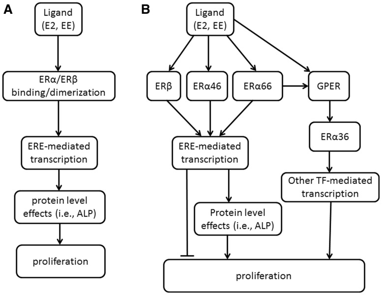
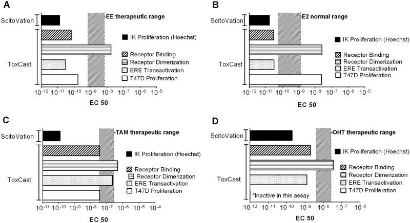
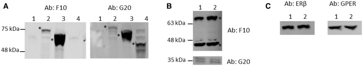
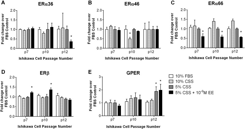
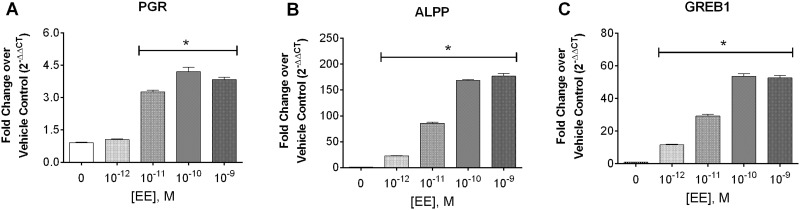
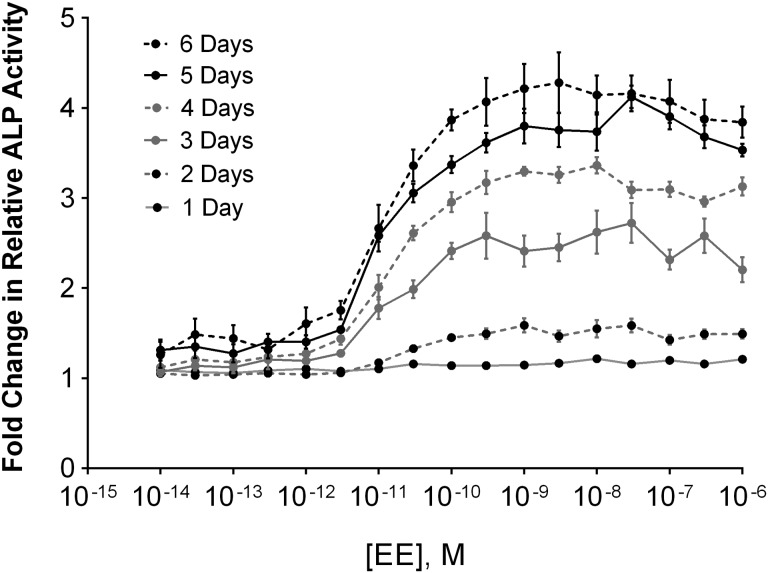
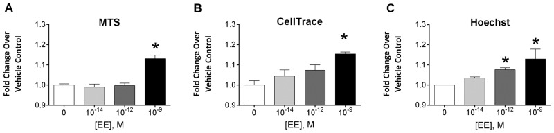
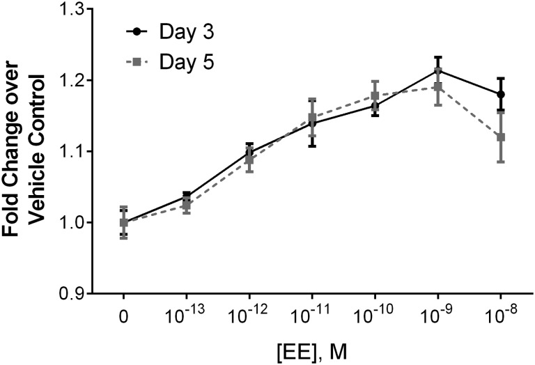
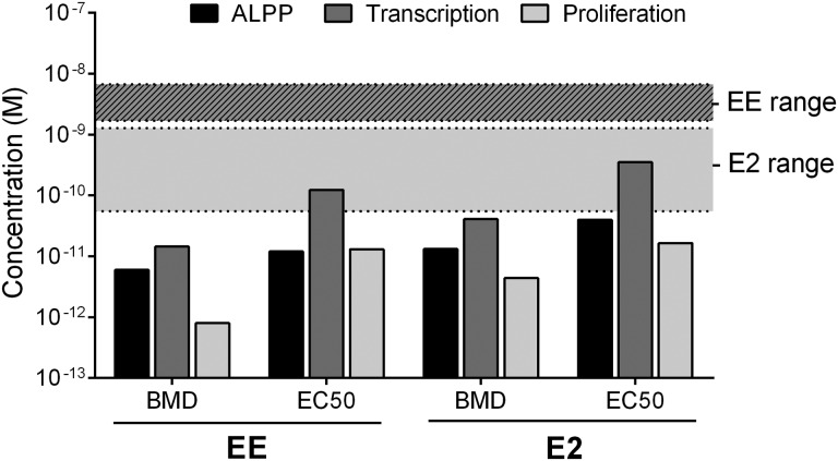
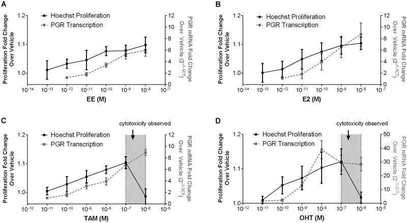
A toxicity pathway approach was taken to develop an in vitro assay using human uterine epithelial adenocarcinoma (Ishikawa) cells as a replacement for measuring an in vivo uterotrophic response to estrogens. The Ishikawa cell was determined to be fit for the purpose of recapitulating in vivo uterine response by verifying fidelity of the biological pathway components and the dose-response predictions to women of child-bearing age. Expression of the suite of estrogen receptors that control uterine proliferation (ERα66, ERα46, ERα36, ERβ, G-protein coupled estrogen receptor (GPER)) were confirmed across passages and treatment conditions. Phenotypic responses to ethinyl estradiol (EE) from transcriptional activation of ER-mediated genes, to ALP enzyme induction and cellular proliferation occurred at concentrations consistent with estrogenic activity in adult women (low picomolar). To confirm utility of this model to predict concentration-response for uterine proliferation with xenobiotics, we tested the concentration-response for compounds with known uterine estrogenic activity in humans and compared the results to assays from the ToxCast and Tox21 suite of estrogen assays. The Ishikawa proliferation assay was consistent with in vivo responses and was a more sensitive measure of uterine response. Because this assay was constructed by first mapping the key molecular events for cellular response, and then ensuring that the assay incorporated these events, the resulting cellular assay should be a reliable tool for identifying estrogenic compounds and may provide improved quantitation of chemical concentration response for in vitro-based safety assessments.
采用毒性途径方法,开发了一种体外试验,使用人子宫上皮腺癌(石川)细胞替代测量雌激素的体内子宫营养反应。通过验证生物途径成分的保真度以及对育龄妇女的剂量反应预测,确定石川细胞适合用于概括体内子宫反应。在不同传代和处理条件下,证实了控制子宫增殖的一系列雌激素受体(ERα66、ERα46、ERα36、ERβ、G蛋白偶联雌激素受体(GPER))的表达。对乙炔雌二醇(EE)的表型反应,从ER介导基因的转录激活到碱性磷酸酶(ALP)酶诱导和细胞增殖,在与成年女性雌激素活性一致的浓度(低皮摩尔)下发生。为了证实该模型预测外源性物质对子宫增殖的浓度反应的效用,我们测试了具有已知人体子宫雌激素活性的化合物的浓度反应,并将结果与ToxCast和Tox21雌激素检测套件的检测结果进行比较。石川细胞增殖试验与体内反应一致,是一种更敏感地测量子宫反应的方法。由于该试验首先通过绘制细胞反应的关键分子事件,然后确保试验纳入这些事件构建而成,因此所得的细胞试验应该是鉴定雌激素化合物的可靠工具,并且可能为基于体外的安全性评估提供改进的化学浓度反应定量。