School of Electrical, Computer and Energy Engineering, Arizona State University, Tempe, Arizona.
Center for Biosignatures Discovery Automation, Biodesign Institute, Tempe, Arizona.
Sci Rep. 2016 Aug 9;6:30593. doi: 10.1038/srep30593.
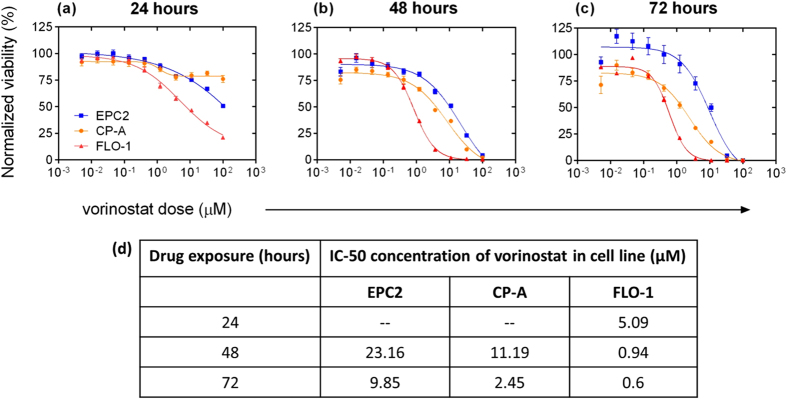
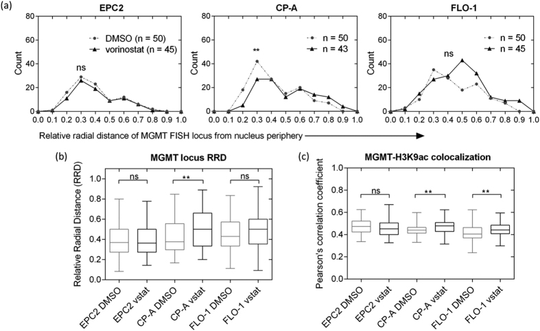
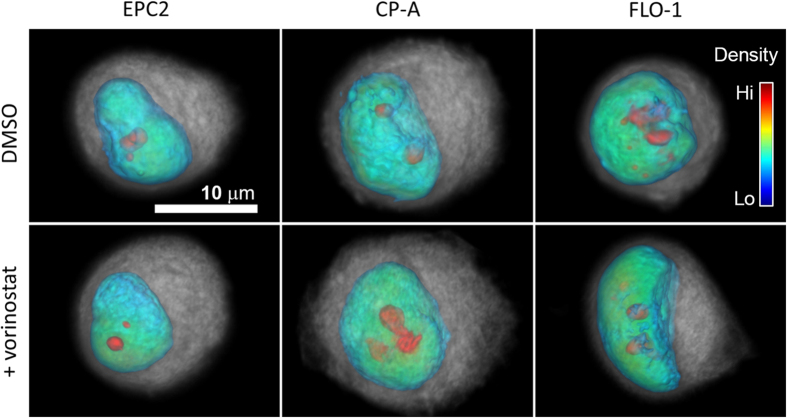
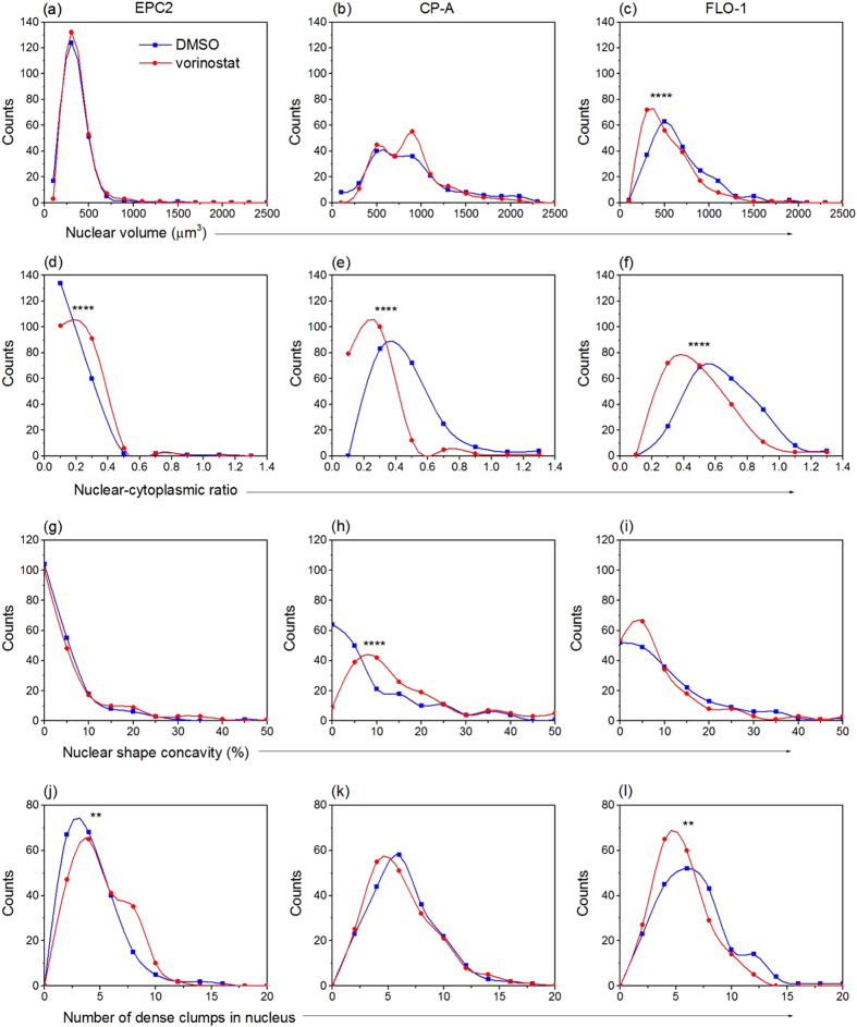
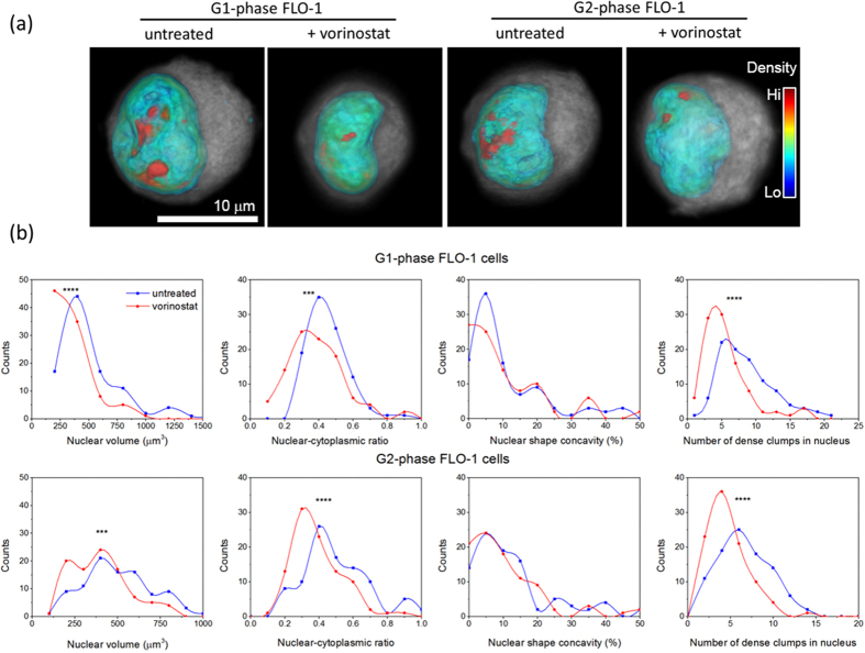
The histone deacetylase (HDAC) inhibitor vorinostat has received significant attention in recent years as an 'epigenetic' drug used to treat solid tumors. However, its mechanisms of action are not entirely understood, particularly with regard to its interaction with the aberrations in 3D nuclear structure that accompany neoplastic progression. We investigated the impact of vorinostat on human esophageal epithelial cell lines derived from normal, metaplastic (pre-cancerous), and malignant tissue. Using a combination of novel optical computed tomography (CT)-based quantitative 3D absorption microscopy and conventional confocal fluorescence microscopy, we show that subjecting malignant cells to vorinostat preferentially alters their 3D nuclear architecture relative to non-cancerous cells. Optical CT (cell CT) imaging of fixed single cells showed that drug-treated cancer cells exhibit significant alterations in nuclear morphometry. Confocal microscopy revealed that vorinostat caused changes in the distribution of H3K9ac-marked euchromatin and H3K9me3-marked constitutive heterochromatin. Additionally, 3D immuno-FISH showed that drug-induced expression of the DNA repair gene MGMT was accompanied by spatial relocation toward the center of the nucleus in the nuclei of metaplastic but not in non-neoplastic cells. Our data suggest that vorinostat's differential modulation of 3D nuclear architecture in normal and abnormal cells could play a functional role in its anti-cancer action.
组蛋白去乙酰化酶(HDAC)抑制剂伏立诺他近年来作为一种“表观遗传”药物受到了广泛关注,用于治疗实体瘤。然而,其作用机制尚不完全清楚,特别是其与肿瘤进展过程中伴随的 3D 核结构异常的相互作用。我们研究了伏立诺他对源自正常、化生(癌前)和恶性组织的人食管上皮细胞系的影响。我们采用新型光学计算断层扫描(CT)基于定量 3D 吸收显微镜和传统共聚焦荧光显微镜相结合的方法,表明将恶性细胞暴露于伏立诺他会优先改变其相对于非癌细胞的 3D 核结构。固定单细胞的光学 CT(细胞 CT)成像表明,药物处理的癌细胞表现出核形态计量的显著改变。共聚焦显微镜显示,伏立诺他导致 H3K9ac 标记的常染色质和 H3K9me3 标记的组成型异染色质的分布发生变化。此外,3D 免疫荧光原位杂交显示,药物诱导的 DNA 修复基因 MGMT 的表达伴随着空间重定位到化生但非非肿瘤细胞的核中心。我们的数据表明,伏立诺他对正常和异常细胞 3D 核结构的差异调节可能在其抗癌作用中发挥功能作用。