Wang Guixin, Wang Jia, Sun Dawei, Xin Jingyi, Wang Liping, Huang Dong, Wu Weichi, Xian Cory J
Department of Orthopaedic Traumatology, Tianjin Hospital, Tianjin, China (mainland).
Department of Orthopedics & Microsurgery, Guangdong No. 2 Provincial People's Hospital, Guanghzou, China (mainland).
Med Sci Monit. 2016 Aug 23;22:2962-71. doi: 10.12659/msm.899485.
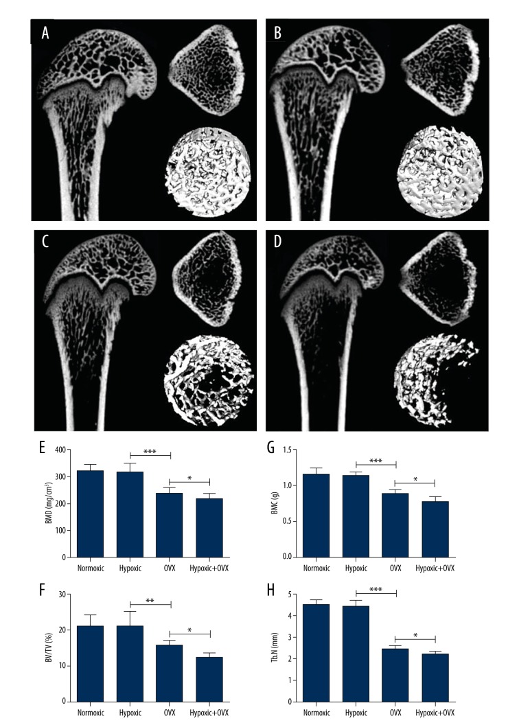
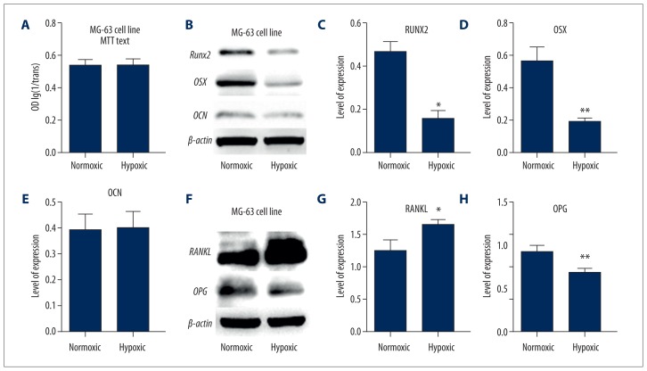
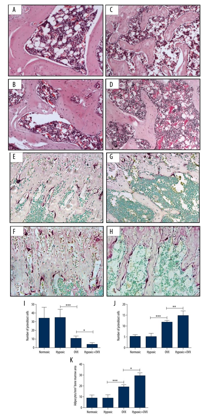
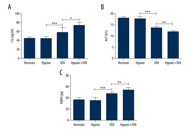
BACKGROUND Although it has been reported that hypoxic exposure can attenuate hypertension, heart disease, diabetes, and some other diseases, effects of hypoxia on osteoporosis are still unknown. MATERIAL AND METHODS The current study investigated whether short-term hypoxic exposure (in comparison with normoxic conditions) affects bone metabolism in normal or ovariectomized (OVX) adult female rats in an vivo study. Micro-computed tomography bone volume/structural analyses, histological examination, and serum bone turnover biochemical assays were used. In addition, the expressions of some associated major regulatory molecules were measured in osteoblastic cultures. RESULTS While the 14-day hypoxic exposure did not change the bone-remodeling process in normal adult female rats, it decreased bone volume, osteoclast density, and serum bone formation marker (alkaline phosphatase) level, but increased osteoclast density and serum bone resorption marker (C-telopeptide of collagen) level in OVX rats. The bone marrow adipocyte number and serum fatty acid binding protein-4 level were increased in OVX-hypoxic rats compared with OVX-normoxic rats. Consistently, in human MG-63 osteoblastic cultures, the hypoxic condition suppressed protein expression of osteogenic transcriptional factors Runx2 and osterix, elevated protein expression of osteoclastogenic cytokine receptor activator of nuclear factor kappa-B ligand, but reduced that of osteoclastogenic inhibitor osteoprotegerin. CONCLUSIONS Our results suggest that, although no change occurred in the bone-remodeling process in normal adult female rats after hypoxic exposure, under the estrogen-deficient osteoporotic condition, the hypoxic condition can alter the bone microenvironment so that it may further impair osteoblastic differentiation and enhance osteoclastic formation, and thus reduce bone formation, enhance bone resorption, and accelerate bone loss.
背景 尽管有报道称低氧暴露可减轻高血压、心脏病、糖尿病和其他一些疾病,但低氧对骨质疏松症的影响仍不清楚。材料与方法 本研究在一项体内研究中,调查了短期低氧暴露(与常氧条件相比)是否会影响正常或去卵巢(OVX)成年雌性大鼠的骨代谢。采用了微型计算机断层扫描骨体积/结构分析、组织学检查和血清骨转换生化测定。此外,还在成骨细胞培养物中测量了一些相关主要调节分子的表达。结果 虽然14天的低氧暴露未改变正常成年雌性大鼠的骨重塑过程,但它降低了去卵巢大鼠的骨体积、破骨细胞密度和血清骨形成标志物(碱性磷酸酶)水平,但增加了破骨细胞密度和血清骨吸收标志物(胶原C端肽)水平。与去卵巢常氧大鼠相比,去卵巢低氧大鼠的骨髓脂肪细胞数量和血清脂肪酸结合蛋白-4水平增加。同样,在人MG-63成骨细胞培养物中,低氧条件抑制了成骨转录因子Runx2和osterix的蛋白表达,升高了破骨细胞生成细胞因子核因子κB配体受体激活剂的蛋白表达,但降低了破骨细胞生成抑制剂骨保护素的蛋白表达。结论 我们的结果表明,虽然低氧暴露后正常成年雌性大鼠的骨重塑过程没有变化,但在雌激素缺乏的骨质疏松条件下,低氧条件可改变骨微环境,从而可能进一步损害成骨细胞分化并增强破骨细胞形成,进而减少骨形成、增强骨吸收并加速骨质流失。