Honnorat Estelle, Seng Piseth, Riberi Alberto, Habib Gilbert, Stein Andreas
Assistance Publique - Hôpitaux de Marseille (APHM), Service de Maladies Infectieuses, Hôpital de la Conception, 147, boulevard Baille, Marseille, France.
Aix Marseille Univ, INSERM 1095, CNRS 7278, IRD 198, URMITE, Marseille, France.
BMC Res Notes. 2016 Aug 24;9(1):416. doi: 10.1186/s13104-016-2223-z.
In contrast to percutaneous atrial septal occluder device, surgical patch closure of atrial defects was known to be no infective endocarditis risk.
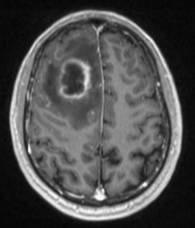
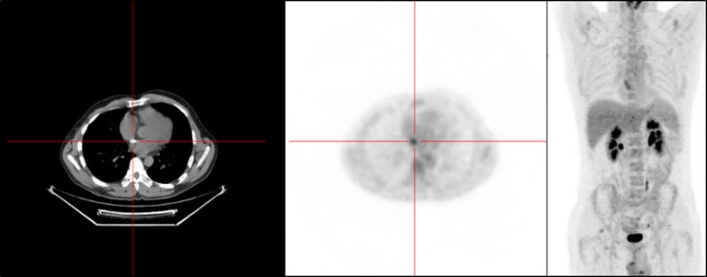
We herein report the first case of late endocarditis of surgical patch closure of atrial septal defects occurred at 47-year after surgery. On September 2014, a 56-year-old immunocompetent French Caucasian man was admitted into the Emergency Department for 3-week history of headache, acute decrease of psychomotor performance and fever at 40 °C. The diagnosis has been evoked during his admission for the management of a brain abscess and confirmed using 18F-fluorodeoxyglucose gated cardiac computed tomography (18F-FDG-PET/CT). Bacterial cultures of surgical deep samples of brain abscess were positive for Streptococcus intermedius and Aggregatibacter aphrophilus as identified by the matrix-assisted laser desorption/ionization-time of flight (MALDI-TOF) mass spectrometry and confirmed with 16S rRNA gene sequencing. The patient was treated by antibiotics for 8 weeks and surgical patch closure removal.
In summary, late endocarditis on surgical patch and on percutaneous atrial septal occluder device of atrial septal defects is rare. Cardiac imaging by the 18F-fluorodeoxyglucose gated cardiac computed tomography (18F-FDG-PET/CT) could improve the diagnosis and care endocarditis on surgical patch closure of atrial septal defects while transthoracic and transesophageal echocardiography remained difficult to interpret.
与经皮房间隔封堵器不同,已知房间隔缺损的外科补片闭合术不存在感染性心内膜炎风险。
我们在此报告首例房间隔缺损外科补片闭合术后47年发生的晚期心内膜炎病例。2014年9月,一名56岁免疫功能正常的法国白种男性因头痛3周、精神运动功能急性减退及40℃发热入住急诊科。入院时考虑脑脓肿诊断并通过18F-氟脱氧葡萄糖门控心脏计算机断层扫描(18F-FDG-PET/CT)得以确诊。脑脓肿手术深部样本的细菌培养经基质辅助激光解吸电离飞行时间质谱(MALDI-TOF)鉴定为中间型链球菌和嗜沫聚集杆菌阳性,并经16S rRNA基因测序确认。患者接受了8周抗生素治疗及外科补片闭合移除术。
总之,房间隔缺损外科补片及经皮房间隔封堵器的晚期心内膜炎罕见。18F-氟脱氧葡萄糖门控心脏计算机断层扫描(FDG-PET/CT)心脏成像可改善房间隔缺损外科补片闭合术心内膜炎的诊断与治疗,而经胸和经食管超声心动图仍难以解读。