Johannsen Finn, Hansen Philip, Stallknecht Sandra, Rathleff Michael Skovdal, Hangaard Stine, Nybing Janus Damm, Boesen Mikael
Institute of Sports Medicine Copenhagen, Bispebjerg Hospital, Building 8, 1., Bispebjerg Bakke 23, Copenhagen, DK-2400, Denmark.
Department of Radiology, Copenhagen University Hospital Bispebjerg & Frederiksberg, Nordre Fasanvej 57, vej 4, opg. 5, Frederiksberg, DK-2000 Denmark.
J Foot Ankle Res. 2016 Sep 1;9(1):35. doi: 10.1186/s13047-016-0168-z. eCollection 2016.
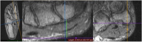
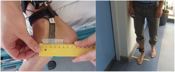
Positional MRI (pMRI) allows for three-dimensional visual assessment of navicular position. In this exploratory pilot study pMRI was validated against a stretch sensor device, which measures movement of the medial plantar arch. We hypothesized that a combined pMRI measure incorporating both vertical and medial displacement of the navicular bone induced by loading would be correlated with corresponding stretch sensor measurements.
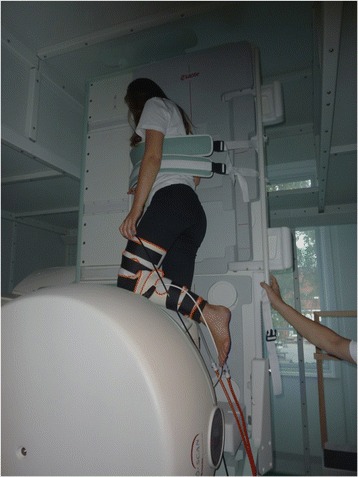
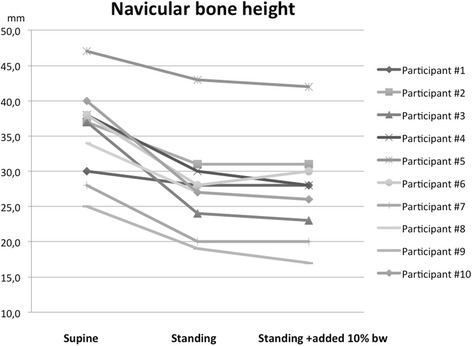
10 voluntary participants were included in the study. Both pMRI and subsequent stretch sensor measurements were performed in a) supine, b) standing and c) standing position with addition of 10 % body weight during static loading of the foot. Stretch sensor measurements were also performed during barefoot walking.
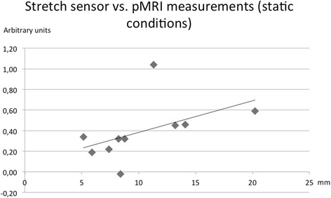
The total change in navicular position measured by pMRI was 10.3 mm (CI: 7.0 to 13.5 mm). No further displacement occurred when adding 10 % bodyweight (mean difference: 0.7 mm (CI: -0.7 to 2.0 mm), P = 0.29). The total navicular displacement correlated with stretch sensor measurement under static loading conditions (Spearman's rho = 0.66, P = 0.04) but not with measurements during walking (Spearman's rho = 0.58, P = 0.08).
Total navicular bone displacements determined by pMRI showed concurrent validity with stretch sensor measurements but only so under static loading conditions. Although assessment of total navicular displacement by combining concomitant vertical and medial navicular bone movements would appear advantageous compared to monoplanar measurement the combined measure did not seem to predict dynamic changes of the medial foot arch during walking, which are among several possible factors depending on different walking patterns.
位置MRI(pMRI)可对舟骨位置进行三维视觉评估。在这项探索性初步研究中,对pMRI与一种测量内侧足弓运动的拉伸传感器装置进行了验证。我们假设,结合负重引起的舟骨垂直和内侧位移的pMRI综合测量值将与相应的拉伸传感器测量值相关。
10名志愿者参与了该研究。pMRI及随后的拉伸传感器测量分别在以下三种状态下进行:a)仰卧位;b)站立位;c)站立位并在足部静态负重时额外增加10%体重。还在赤脚行走过程中进行了拉伸传感器测量。
通过pMRI测量的舟骨位置总变化为10.3毫米(置信区间:7.0至13.5毫米)。增加10%体重后未出现进一步位移(平均差异:0.7毫米(置信区间:-0.7至2.0毫米),P = 0.29)。在静态负重条件下,舟骨总位移与拉伸传感器测量值相关(斯皮尔曼等级相关系数ρ = 0.66,P = 0.04),但与行走过程中的测量值无关(斯皮尔曼等级相关系数ρ = 0.58,P = 0.08)。
pMRI测定的舟骨总位移与拉伸传感器测量值具有同时效度,但仅在静态负重条件下如此。尽管与单平面测量相比,通过结合舟骨垂直和内侧运动来评估舟骨总位移似乎具有优势,但该综合测量似乎无法预测行走过程中内侧足弓的动态变化,而行走过程中的动态变化是取决于不同行走模式的几个可能因素之一。